Mechanisms and Regulation of pre-mRNA Splicing

Professor Vlad Pena's group focuses on the structural basis of human pre-mRNA processing under normal, pathological and drug-induced conditions.

Research, projects and publications in this group

Our group focuses on the structural basis of human pre-mRNA processing, including: splicing modulation by compounds with antitumour properties,  ubiquitination in splicing and DNA repair and helicases.

Catalytic activation of human spliceosomes by RNA helicases

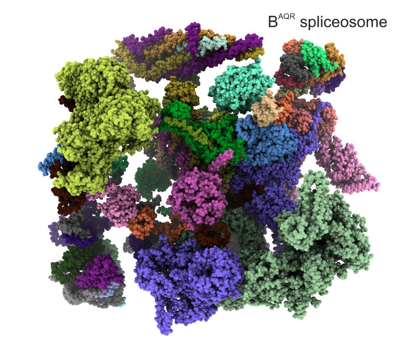
RNA helicases are molecular motors that convert the energy stored in ATP into various rearrangements of RNA and RNA-protein complexes, keeping the RNA metabolism dynamic and tightly regulated. Spliceosomes, which are the largest and most complex nanomachines that process genetic information, require numerous helicases to regulate their function. Using cryo-EM of stalled spliceosome and biochemical methods, we found that human spliceosomes need to be remodelled by two helicases – PRP2 and Aquarius, to kick-start the splicing reaction. 


Spliceosome stalled halfway through the catalytic activation

Identification of a spliceosome stalled halfway through the catalytic activation (Schmitzova et al., Nature, 2023).


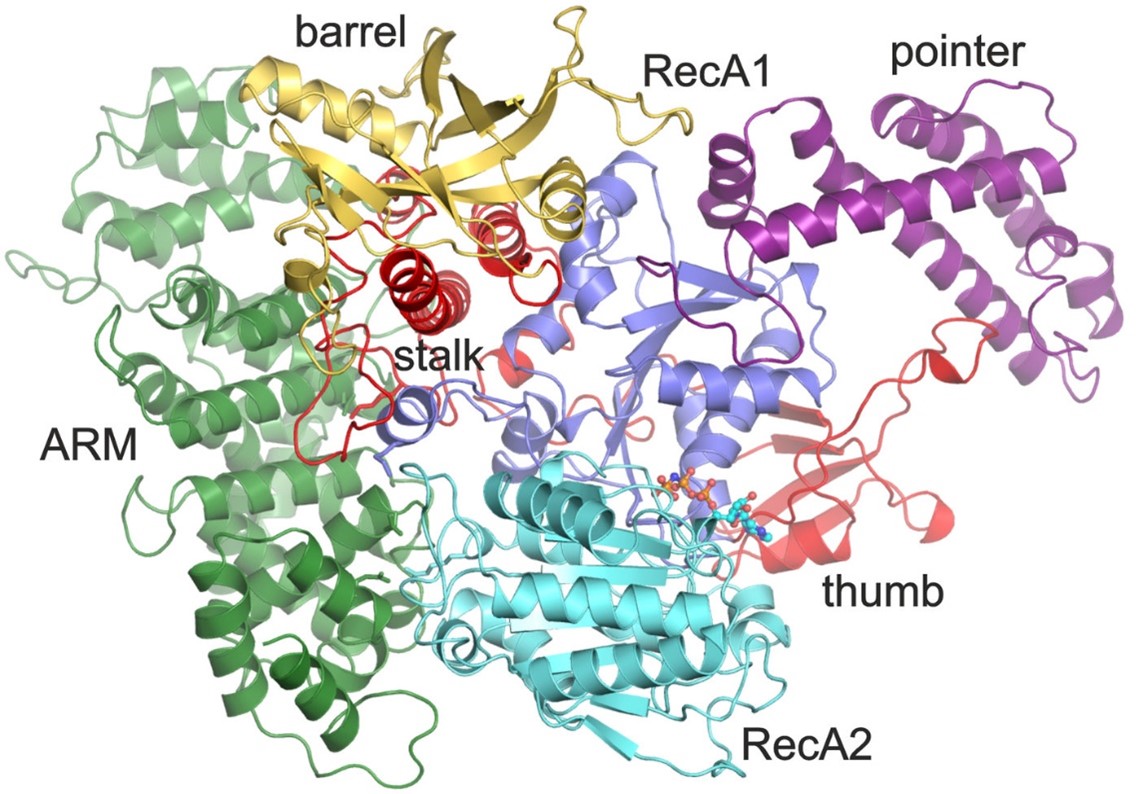
Mechanism of the helicase PRP2 in splicing 

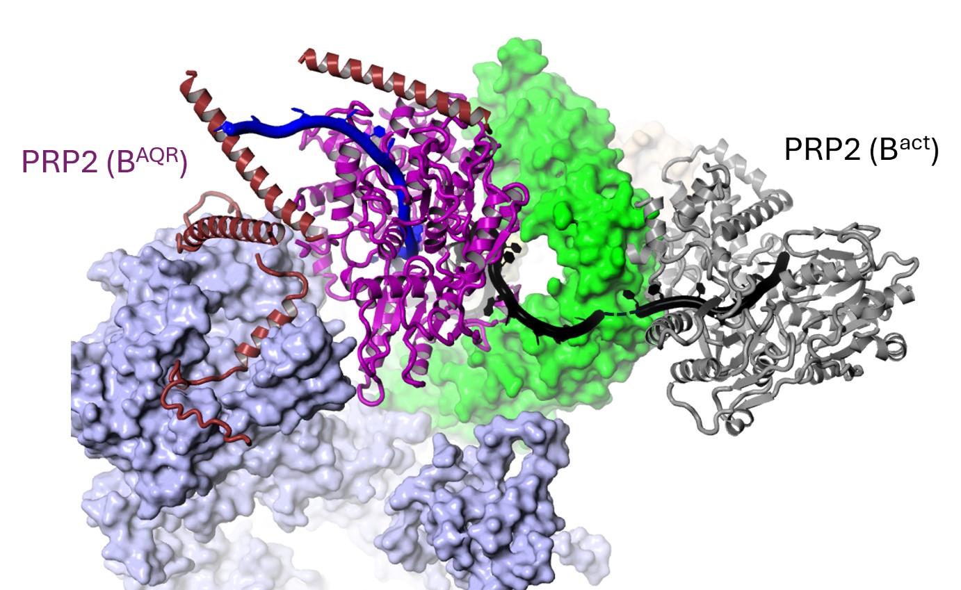
The DEAH helicases are special as they can grasp an RNA strand and translocate along nucleotide by nucleotide while hydrolyzing ATP molecules. Four DEAH helicases are essential for splicing, although their mechanism of action in splicing remained unclear, as they were observed at fixed positions on the periphery of the spliceosomes. The BAQR spliceosome, found halfway through the catalytic activation, has finally shown how a DEAH helicase, PRP2, translocates about 19 nucleotides from the periphery towards the core of the spliceosome while remodelling the RNA-protein contacts, changing conformations, dissociating and recruiting proteins. 


Helicase PRP2

The helicase PRP2 translocates 19 nucleotides at the transition from Bact to BAQR (Schmitzova et al., Nature, 2023).


General mechanism of intron recognition during spliceosome assembly

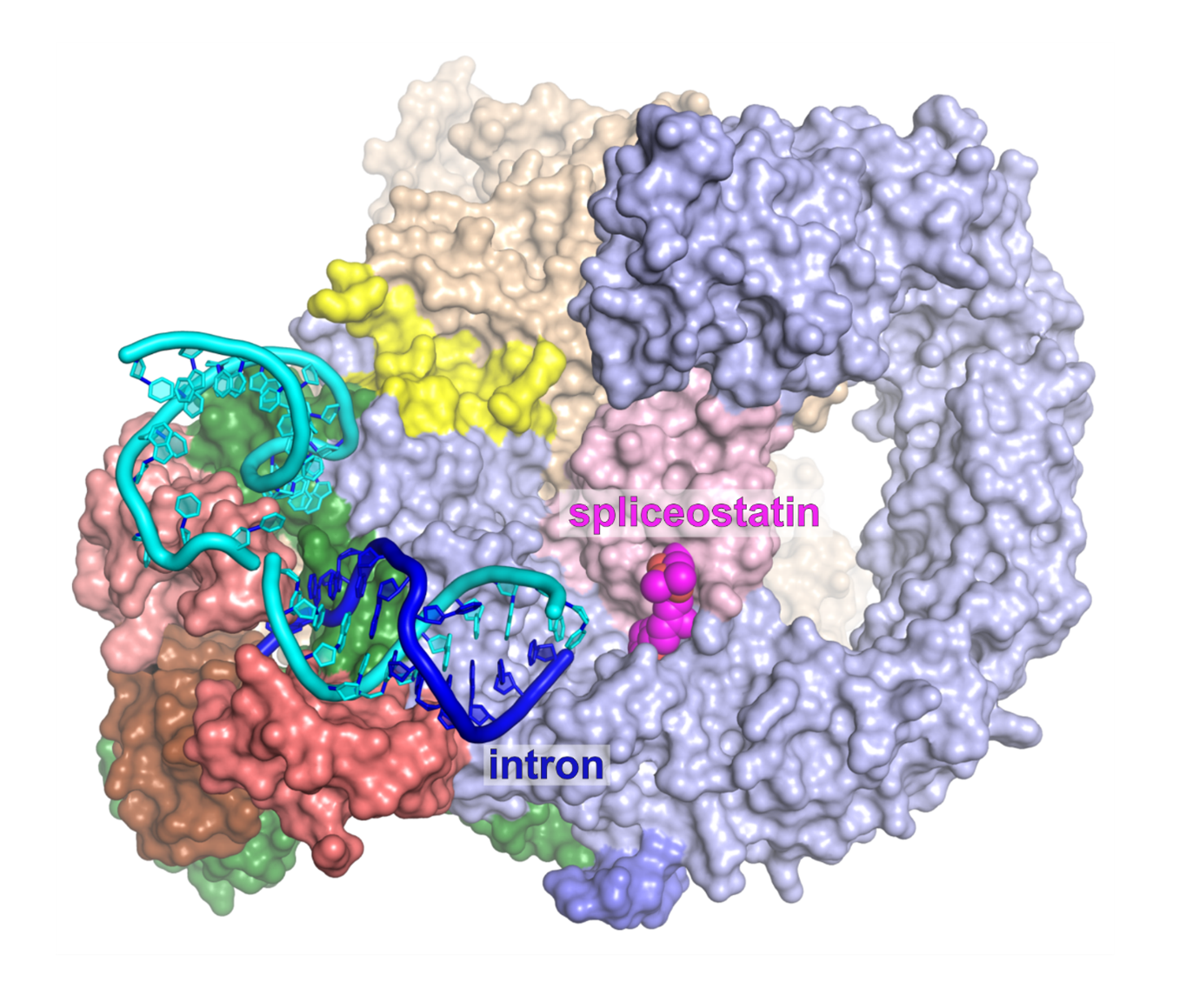
The structure of a cross-exon spliceosome stalled by SSA captures the intron recognition by U2 snRNP.
This indicates that early prespliceosomes (E complexes) select introns through a toehold-mediated strand displacement mechanism. Strand displacement is primarily known from the DNA field, on processes like the DNA strand exchange, and in applied DNA nanotechnologies. 


Exon-definition complex coreIntron recognition by strand displacement

The structure of the exon-definition complex core stalled with spliceostatin (above) and the mechanism of intron recognition by strand displacement (below; Cretu et al., Nat. Comm., 2021).


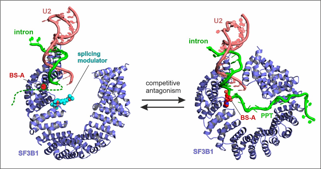
General mechanism of splicing modulation by SF3B ligands

SF3B ligands with antitumor properties act as competitive antagonists of branch sites (see SF3B-PB, SF3B-SSA, SF3B-SUD). Our structures explain why the inhibition depends on the sequence motifs of the intron, as such that some branch sites can be inactivated more readily than others. The modulation manifests as differential inhibitions (some branch sites will be used while some others not), resulting in a change in AS pattern (Cretu et al., Mol. Cell, 2018; Nat Commun 2021).


Splicing modulators

Splicing modulators that bind SF3B1 act as competitive antagonists of the branch site, in a manner dependent on the intron’s sequence (Cretu et al., Mol. Cell, 2018; Nat. Comm., 2021)


Mechanism of covalent coupling of spliceostatin and sudemycine to a zinc finger of SF3B

Our crystal structures have revealed that the long-studied compounds from the spliceostatin family and their relatives, sudemycins, bind prespliceosomes by covalent coupling to a zinc finger of the PHF5A protein. The central conjugated diene of the compounds acts as a spacer for the warhead epoxy group that reacts with a thiol of the cysteine. 

Spliceostatin and sudemycins bind spliceosomes

Spliceostatin and sudemycins bind spliceosomes by covalent coupling to a zing finger from the SF3B subunit PHF5A.


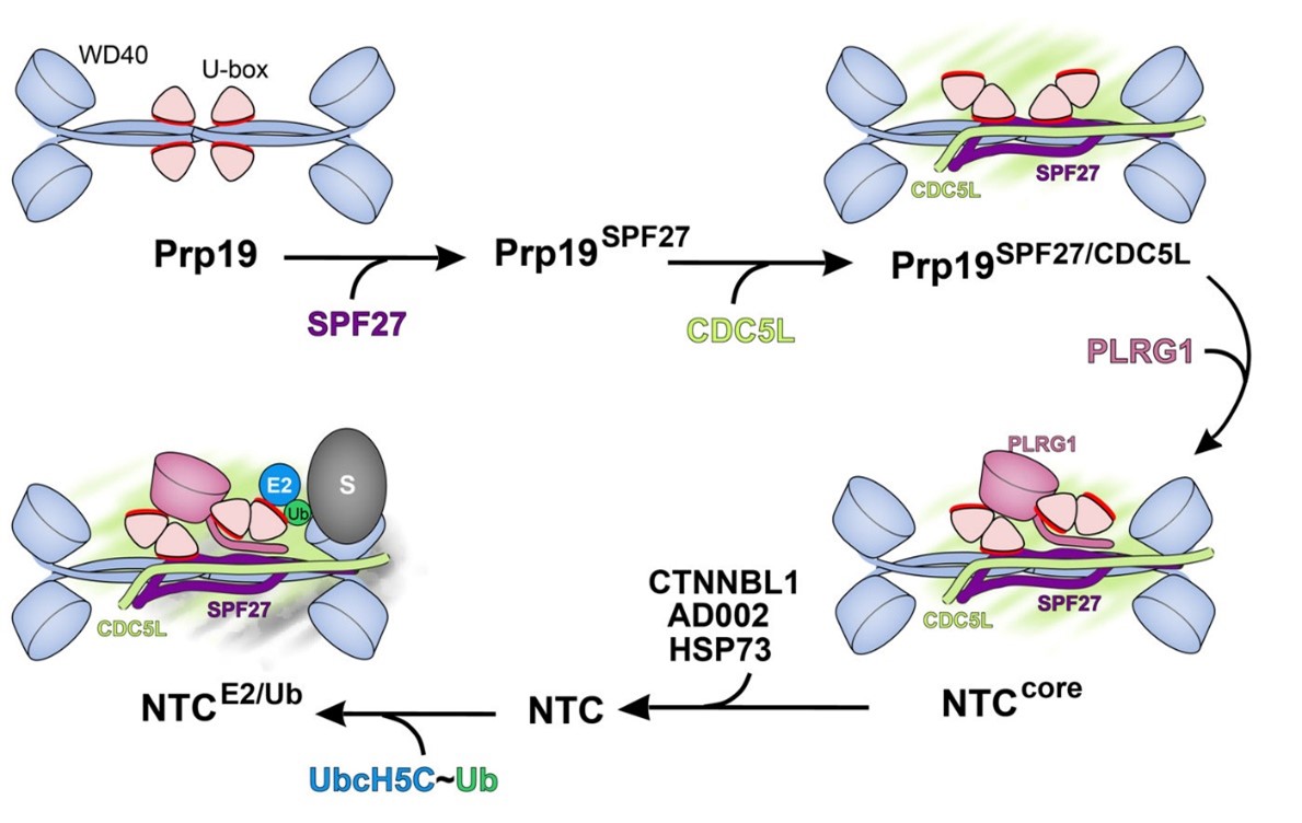
The NineTeen Complex (NTC) promotes ubiquitination in DNA repair

As part of the heteromeric NineTeen Complex (NTC), Prp19 is a homotetramer complex that promotes ubiquitination during the formation of the spliceosomal tri-snRNP particle and DNA damage response. We showed by X-ray crystallography and functional analyses in vitro and in vivo that Prp19 is an autoinhibited E3 ubiquitin ligase that becomes active only in stable association with three other NTC components that exert specific effects on Prp19's conformation. (De Moura et al., Mol. Cell, 2018).


PRP19 activated by stepwise assembly of splicing factors

PRP19 is an autoinhibited E3 ligase activated by stepwise assembly of three splicing factors, together forming the NTC’s core


The Intron-binding complex (IBC) enables recruitment of the splicing-essential helicase Aquarius (AQR, IBP160) to the human spliceosomes

The human helicase Aquarius induces an additional ATP-dependent remodeling of the spliceosome. We determined the structure of Aquarius and showed that this helicase is recruited to the spliceosome as part of the pentameric intron binding complex (IBC).


Crystal structure of the RNA helicase Aquarius


Crystal structure of the RNA helicase Aquarius


Crystal structure of a DNA catalyst

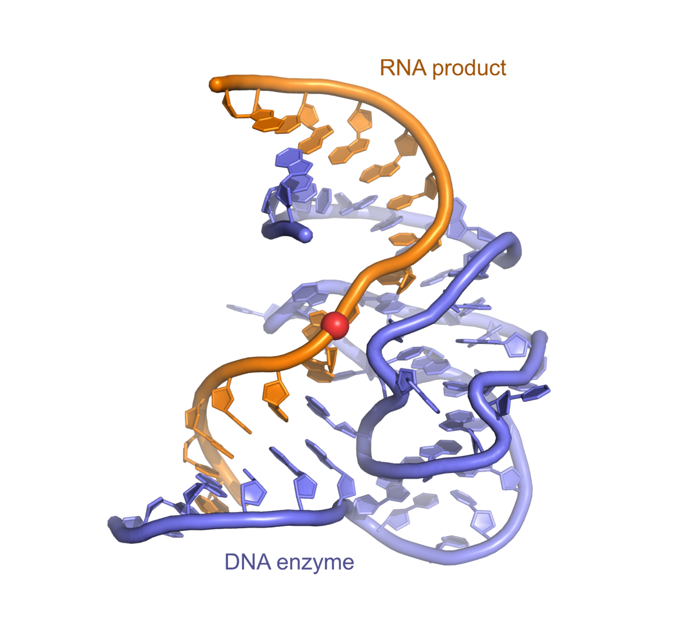
Although DNA is known mainly for its capacity to encode genetic information, DNA molecules that catalyze various chemical reactions have been found in vitro. Two decades after this discovery, we have determined the first crystal structure of a DNA enzyme. The surprisingly complex fold that these molecules adopt raises questions about the potential structural importance of DNA in the cell (Ponce-Salvatierra et al., Nature 2016).


deoxyribozyme 9DB1 in complex with the RNA product

Crystal structure of the deoxyribozyme 9DB1 in complex with the RNA product

Professor Vlad Pena

Group Leader:

Mechanisms and Regulation of pre-mRNA Splicing Vlad Pena

Professor Vlad Pena uses biochemistry, electron cryo-microscopy and X-ray crystallography to understand splicing regulation and the connections between splicing and other gene expression processes. He is a Professor of Structural Biology and Gene Expression at the ICR.

Researchers in this group

Portrait of Imogen Andrews, smiling .

Email: [email protected]

Location: Chelsea

Imogen is a PhD student in the Division of Structural Biology focused on designing small molecule splicing modulators, in collaboration with the Centre for Cancer Drug Discovery.

Tiffany Bergot profile picture .

Phone: +44 20 7153 5524

Email: [email protected]

Location: Chelsea

Tiffany is a postdoctoral fellow who works on SF3B functions relevant to cancer.

Judit Galicza profile picture .

Email: [email protected]

Location: Chelsea

Judit is a higher scientific officer studying the regulation of RNA processing.

Negar Goodarzi profile picture .

Phone: +44 20 3437 6398

Email: [email protected]

Location: Chelsea

Gus explores the modulation of gene expression by oncolytic viruses.

Csaba Kibedi profile picture .

Email: [email protected]

Location: Chelsea

Csaba is a postdoctoral fellow working on splicing regulation.

Hyunah Lee profile picture .

Phone: +44 20 7153 5498

Email: [email protected]

Location: Chelsea

Hyunah is a postdoctoral fellow who studies the regulation of alternative splicing.

Ioana Maruntel profile picture .

Phone: +44 20 7153 5090

Email: [email protected]

Location: Chelsea

.

OrcID: 0000-0002-3975-5932

Email: [email protected]

Location: Chelsea

I am a PhD student and I’m interested in understanding the functions and structure of the splicing molecular machinery and capture its linking to other close related events in gene expression.

Professor Vlad Pena's group have written 24 publications

Most recent new publication 7/2023

See all their publications

News and discoveries from this group

10/05/23

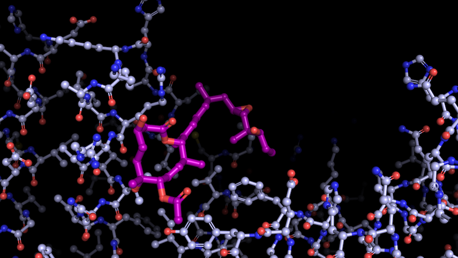
This represents a splicing modulator bound to a spliceosomal component

Image: “Like a constellation of atoms” – splicing modulator (purple) bound to a spliceosome subunit. Credit: Professor Vlad Pena. 

Scientists have uncovered the inner workings of one of the most important and intricate ‘nanobots’ operating within our cells – using cutting-edge microscopy for visualising molecules almost at an atomic level.

Their new study published in Nature has unveiled the critical step that switches on the spliceosome – a piece of cellular machinery that enables cells to build complex proteins.

By uncovering in detail how the spliceosome is activated, scientists believe the discovery could pave the way to more effective design of cancer drugs which target it.

State of the art microscopy

An international team of scientists from The Institute of Cancer Research, London, and the Max Planck Institute for Multidisciplinary Sciences in Germany employed state-of-the-art biochemical and cryo-electron microscopy (cryo-EM) methods to study the spliceosome in intricate detail and answer long standing questions about how it works.

The spliceosome operates like a nanobot, processing RNA – genetic instructions copied from DNA – in a key step to allow the building of complex proteins.

Powered by molecular motors called helicases, the spliceosome chops and changes RNA code to increase the complexity of the genetic instructions so that many different proteins can be made from a limited number of genes. This process is called splicing.

Splicing explains why humans, who only have around 20,000 genes, can produce hundreds of thousands of different proteins. It may also be a key reason for why humans can be so different from fruit flies, despite having a similar number of genes1.

Hallmark of cancer

Mutations in the spliceosome are a hallmark of cancer – they contribute to the production of abnormal proteins that fuel tumour growth or deactivate proteins that protect against cancer. 

Scientists studied the spliceosome using cryo-EM – a cutting edge microscopy technique which involves rapidly freezing spliceosomes and bombarding them with electrons to obtain a 3D reconstruction of their molecular structure at almost atomic-level resolution.

They also employed advanced biochemical engineering methods to capture the spliceosome in the midst of activation – a feat never achieved before. This allowed them to dissect the precise molecular mechanisms occurring within the spliceosome, much like an engineer taking apart an engine but on a sub-microscopic scale. 

Of particular interest was a core spliceosome subunit called SF3B1 which is essential for  spliceosome activation. SF3B1 is the most mutated spliceosome gene in cancer, in particular in leukaemia, uveal melanoma, pancreatic, and prostate cancer. 

The researchers discovered that two molecular motors reshape SF3B1, and in doing so they kickstart splicing.

Firstly, they showed that a molecular motor called PRP2 interacts with SF3B1 and works in a completely new way than was previously thought for splicing helicases. Instead of staying on the outside of the spliceosome, PRP2 “walks” along the RNA strand being processed, all the way to the spliceosome’s core, rearranging the spliceosome structure as it travels and helping to switch the spliceosome into an active state. The researchers believe that other helicases may also work in this new and unexpected way.

Secondly, they found that a second motor named Aquarius also acts on SF3B1 and is essential to activate the spliceosome.

The findings represent a fundamental advance in our understanding of the spliceosome and how it is activated by helicases.

Professor Vlad Pena, Professor of Structural Biology and Gene Expression at The Institute of Cancer Research, London, who supervised the research team said:

“The spliceosome is an intricate nanobot that uses molecular motors to process genetic information. This information is passed on and forms instructions for building proteins.

"We used a new engineering technique to reveal that kickstarting the spliceosome requires the help of two distinct motors, PRP2 and Aquarius. This is a breakthrough finding in our understanding of how the spliceosome and its molecular motors operate.

“Splicing is often dysregulated in cancer, and we hope our work will inspire new research which will contribute to the design of new cancer drugs that can target the splicing process.”

Discovery could pave the way to better cancer drugs

Professor Kristian Helin, Chief Executive of The Institute of Cancer Research, London, said:
 
“These exciting findings represent a fundamental advance in our understanding of one of the most important and complex pieces of molecular machinery in our cells. The spliceosome not only enables complex life to exist but, when things go wrong, it can create proteins which help to fuel or sustain cancer.

“By illuminating the step-by-step process that activates the spliceosome, this research could pave the way to better cancer drugs to control the way it operates within cancer cells."

Help us revolutionise cancer treatment. We aim to discover a new generation of cancer treatments so smart and targeted, that more patients will defeat their cancer.
Donate now