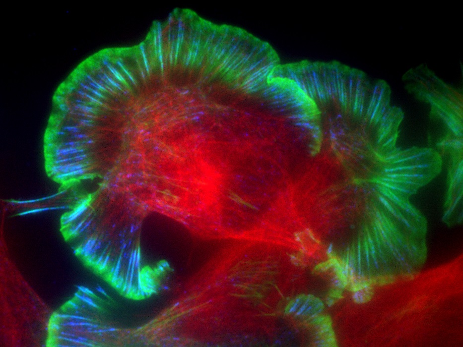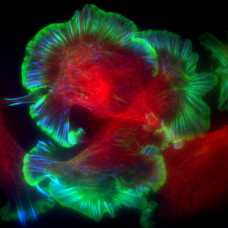
Dr Chris Bakal and Oliver Inge's winning photgraph: Metastatic mouse melanoma cells spreading on a collagen matrix and imaged by Total Internal Reflection Fluorescence (TIRF) microscopy. Actin in green, focal adhesions in blue, and tubulin in red.
Dr Chris Bakal’s and Oliver Inge's image of metastatic melanoma cells – which they described as master shape-shifters – has been crowned the winner of the ICR's 2017 Science Photography and Imaging Competition.
When cancer spreads through the body, metastatic cells make journeys through different environments with radically different compositions – from soft, fluid-like tissues to hard bone. Crossing each tissue requires metastatic cells to assume different shapes.
Sign up to see all of the incredible images below and many more, on display in Sutton Library throughout July.
The winning image shows metastatic melanoma cells as they feel their way through their environment using adhesions (marked in blue) at the plasma membrane of cells, changing their shape to suit the tissue they are invading.
Dr Bakal took this image using the new 'total internal reflection fluorescence' (TIRF) microscope, which allows us to see cells at the point where they are making contact with their environment, and capture structures such as adhesions that would otherwise be difficult to see.
The judging panel selected the image for its visual impact, and for its ability to bring a complicated scientific concept to life. The image was commended for its strong sense of movement suggesting something was about to happen, and for the way the image was related back to the patient.
Dr Bakal and Dr Inge will receive a prize of £250, and the image will be displayed as artwork around the ICR and at the ICR conference later in the year, together with the other shortlisted entries.
Runner-up entries:
Dr Sebastian Guettler
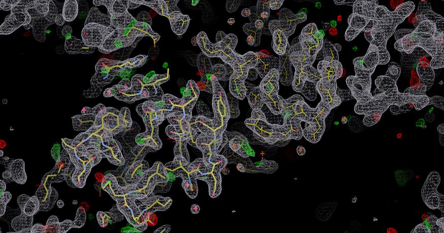
Dr Sebastian Guettler's photograph: Seeing is understanding – crystal structure of the polymer-forming domain of a tankyrase protein.
Dr Sebastian Guettler, leader of the Structural Biology of Cell Signalling Team, used an approach called X-ray crystallography to produce this image. The technique involves collecting data on X-ray diffraction by protein crystals, and using that to produce an electron density map.
This image shows the polymer-forming domain of a protein known as tankyrase – important in cancer signalling. This is the laboratory's first crystal structure, determined by PhD student Laura Mariotti.
Dr Maxine Lam
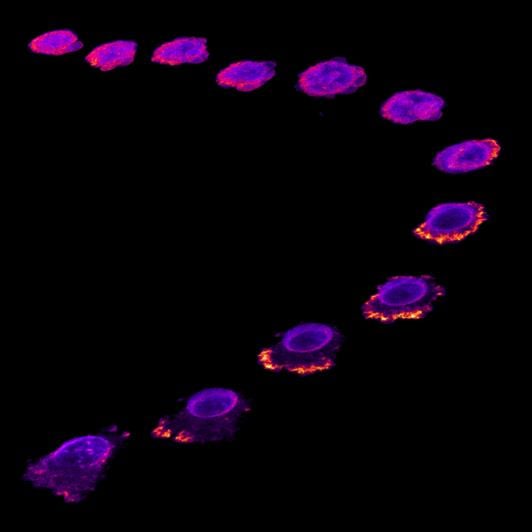
Dr Maxine Lam's photograph: Time lapse image of a breast cancer cell escaping the blood vessel layer during metastasis.
This image depicts a key step in cancer metastasis when cancer cells circulating in the blood stream escape through the endothelial layer of blood vessels. This process is known as extravasation.
It was taken by Dr Maxine Lam, Postdoctoral Training Fellow in the Cancer Biology division, using fluorescence microscopy and captures the process in action.
Breast cancer cells labelled with a fluorescent protein (actin) can be seen exploring and interacting with the endothelial layer, before breaching and crossing through it.
Dr David Mansfield
![Runner up - D Mansfield - 1044_[59009,11249]_composite_image Runner up - D Mansfield - 1044_[59009,11249]_composite_image](/images/default-source/migrated/scientific/runner-up---d-mansfield---1044_-59009-11249-_composite_image.jpg)
Dr David Mansfield's photograph: Tumour cells interacting with their microenvironment to investigate immuno-suppression.
This image was taken by Dr David Mansfield, Higher Scientific Officer in the Radiotherapy and Imaging division, on the ICR's brand new seven-colour fluorescence microscope.
Tumours establish intricate relationships with a host of cells in the body as they grow and invade healthy tissue, and the interaction with immune cells is particularly complex.
Using immunofluorescence imaging, this image shows a tumour in white, surrounded by red, green and yellow lymphocytes, which are types of white blood cells that play a key role in immune reactions. The tumour has been infiltrated by macrophages, in blue.
Some of the green lymphocytes have a magenta nucleus, indicating a suppressive cell is present that prevents other immune cells from attacking. These suppressive cells are a key target for immuno-oncology drugs.
Dr Orli Yogev
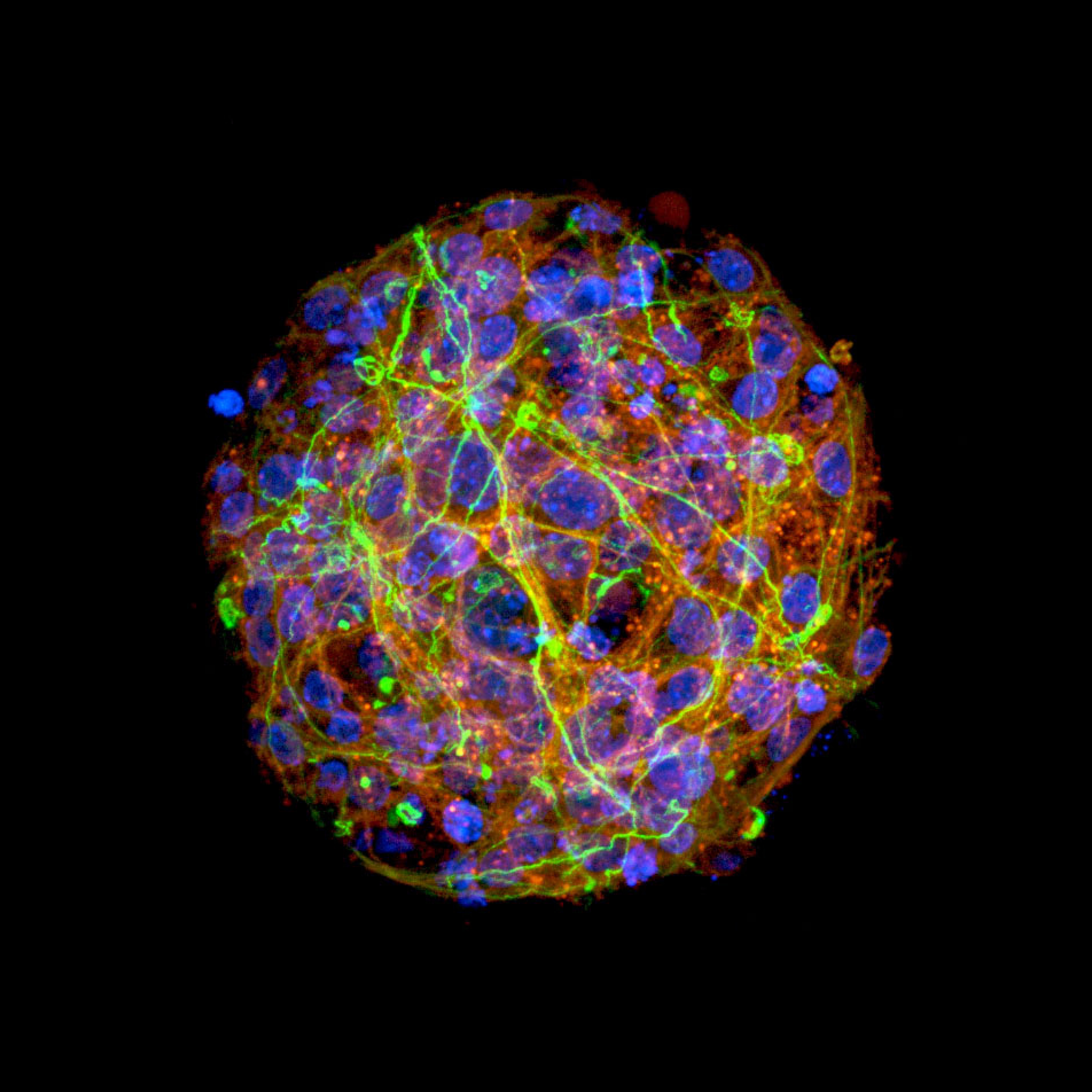
Dr Orli Yogev's photograph: An in vivo model of bone marrow metastasis of neuroblastoma to investigate treatment resistance and metastatic disease.
This image was taken by Dr Orli Yogev, Postdoctoral Training Fellow in the Clinical Studies division, using 3D confocal imaging.
Neuroblastoma is a type of childhood cancer that in its high-risk form is very aggressive, spreads quickly and responds poorly to conventional treatments. By developing tumour models, we can study how cancer responds to chemotherapyand becomes resistant.
This image shows metastatic bone marrow cells (red) of advanced neuroblastoma that has become resistant to chemotherapy. Visible is a subpopulation of neural stem cells (green) that could potentially be a source of metastatic relapse.
The judging panel
The judging panel was made up of:
- Professor Nandita deSouza, Professor of Translational Imaging
- Catherine Graham, Head of Brand and Creative
- Rose Wu, Internal Communications Manager at the ICR
- Dr Nick van As, Consultant Clinical Oncologist
- Ben Hartley, Arts Officer at The Royal Marsden NHS Foundation Trust
- Dr Sabrina Taner, Biomedical Image Acquisitions and Picture Researcher at the Wellcome Trust.
Professor Nandita deSouza said:
“The quality of the images submitted to the ICR's science photography competition was incredibly high, and it was a pleasure to be part of the panel.
“The judges were struck by the strength of the story-telling that went into the images – and how these were used to illustrate the different approaches we use to understand and tackle cancer. Congratulations to everyone who entered.”
Dr Nick Van As said:
“Researchers at the ICR and The Royal Marsden are producing fascinating, informative and beautiful images in their research. The winning image really captured what the judges were looking for – a marriage of visual impact and scientific value.”
