Ultrasound and Optical Imaging Group
Professor Jeffrey Bamber’s group develops new ways of using ultrasound imaging to detect various cancers, including breast, prostate and skin cancer.
Our group’s work aims to invent, develop and apply new ways of deriving information from ultrasound signals and utilising ultrasound waves to assist in cancer treatment.
Our overall interest is in increasing the functional and molecular imaging capability of ultrasound, providing new tools to experimental cancer biology and helping to personalise cancer treatment by bringing the cost-effectiveness, safety, speed, patient acceptability, interactiveness and easy reusability of ultrasound based methods to clinical problems such as diagnosis, assessing tumour aggressiveness and response, and guiding treatment.
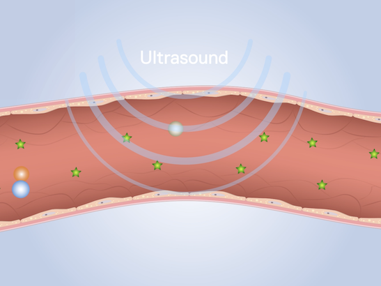
Current research interests in support of these areas aims to develop and apply novel quantitative ultrasound characteristics, biomechanical imaging methods, dynamic tissue tracking, multispectral photoacoustic imaging, quantitative microbubble contrast agent kinetics, photoacoustic probes and methods for using diagnostic devices to locally enhance delivery of drugs and other therapeutics to tumours.
The research interests of the Ultrasound and Optical Imaging Group include:
- High frequency transducers and arrays
- Freehand elastography – breast imaging
- Freehand elastography – neurosurgical guidance
- Freehand elastography – a hybrid 3D strain image acquisition technique
- Quantitative elasticity imaging – elastic modulus and its use for ionising radiation dosimetry
- Quantitative elasticity imaging - porosity and permeability
- Quantitative elasticity imaging – slip elastography
- Quantitative elastography – improving lateral displacement and strain measurement
- High resolution and microscopic elastography
- Organ motion tracking for motion compensated therapy
- Clinical freehand reflection-mode photoacoustic imaging
- Illumination optimisation for freehand reflection-mode photoacoustic imaging
- Photoacoustic absorption spectroscopy and gold nanorods for molecular imaging
- Photoacoustic imaging and emission spectroscopy of tumour vascularisation
- Dynamic contrast-enhanced ultrasound (DCE-US) for tumour response
- Acoustically activated nanoparticle agents for molecular imaging
- Multimodality imaging of apoptosis
We are only able to engage in such diversity via extensive collaboration. In applications to assist radiotherapy, the work of the Ultrasound and Optical Imaging Group is highly integrated with that of the Imaging for Radiotherapy Adaptation team. Strong collaborations also exist with other groups, within the Institute of Cancer Research and Royal Marsden, with other Universities and Colleges, and with industry - notably with Imperial College London, University College London, Queen Mary University of London, The University of Leeds, The University of Glasgow, The University of Edinburgh, iThera GmbH, Delphinus Medical Technologies, Michelson Diagnostics and Phoenix Solutions AS.
Ultrasonic imaging provides essential in-vivo anatomical and functional information that can be used in cancer medicine for early detection, differential diagnosis, staging, biopsy guidance, treatment planning, treatment guidance, and the assessment of response to treatment. In combination with appropriate injectable agents, diagnostic ultrasound may also enhance the delivery of drugs and other therapeutics to tumours, thus increasing their effectiveness and minimising treatment side effects.
Our group’s work aims to enhance these functions by inventing, developing and applying new ways of deriving information from ultrasound signals and utilising ultrasound waves to assist cancer treatment. Optical methods complement ultrasound, with considerable potential for adding new information to that provided by ultrasound as, for example, in the multiphysics imaging method of photoacoustic imaging.
Our research is strongly translational, from basic physics and technical development, through preclinical studies to clinical evaluation. It supports various fields of application, including:
- Breast and breast cancer assessment
- Skin-cancer diagnosis
- Assessment of side-effects of breast-cancer treatment
- Image guidance of treatment (focused-ultrasound, radiotherapy, and surgical resection)
- Prostate-cancer assessment
- The biology of tumour vascularisation and invasion
- Characterisation of tumour phenotype
- Assessment of tumour response.
The group also provides scientific and technical support for activities in the Royal Marsden NHS Foundation Trust. These include assisting with clinical ultrasound research of the Trust, advice to clinical personnel on specific aspects of the safe and effective use of ultrasound, and aiding the purchase of, acceptance testing, quality assurance, acoustic safety testing and first line maintenance of clinical ultrasound equipment.
Professor Jeff Bamber
Group Leader:
Ultrasound and Optical Imaging
Professor Jeffrey Bamber is researching ways to improve the use of ultrasound in spotting tumours. Professor Bamber is past president of the International Association for Breast Ultrasound and past vice-president of the International Society of Skin Imaging.
Researchers in this group
Professor Jeff Bamber's group have written 308 publications
Most recent new publication 4/2025
See all their publicationsIndustrial partnership opportunities with this group
Opportunity: Novel non-invasive imaging tool for measuring tumour stiffness
Commissioner: Dr Emma Harris
Recent discoveries from this group

 .
.


