Magnetic Resonance Imaging in Radiotherapy Group

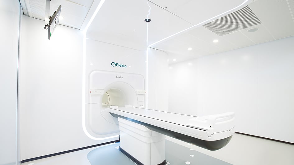
Dr Andreas Wetscherek's group are developing new MRI techniques for radiotherapy with particular application to the UK's first MR-Linac machine.

We develop MRI techniques that enable novel MR-guided radiotherapy workflows.

Background

Radiotherapy has been instrumental in the treatment of cancer for more than 100 years. Treatment is delivered over several fractions to improve tumour control and minimise normal tissue complications. Radiotherapy treatments are planned using electron density information obtained from computed tomography (CT) images to deliver the prescribed dose to the tumour while minimising dose to normal tissue.

Imaging biomarkers that could assess and predict response to treatment are of particular interest, for example to identify resistant subvolumes of the tumour or early signs of side-effects of radiation. MR-guided radiotherapy on a hybrid MR-Linac system enables novel treatment workflows but requires imaging solutions that are different from diagnostic MRI. Our research focuses on two key applications: Functional MRI to assess and predict response to therapy and 4D MRI to manage motion.

Functional MRI

To assess response to treatment, techniques based on diffusion-weighted MRI are particularly promising due to their association with response-related metrics, such as cellularity and cell membrane permeability. Our research interest here is to minimise geometric distortions in diffusion-weighted MRI for use in daily adaptive MR-guided radiotherapy and to enable the simultaneous characterisation of perfusion and diffusion using flow-compensated intravoxel incoherent motion (IVIM) imaging.

In MR-guided radiotherapy with MeV photons, the formation of reactive oxygen species plays an important role in the DNA damage mechanisms and reduced oxygenation (hypoxia) in the tumour is associated with resistance to radiotherapy. We’re working on fast MR relaxometry techniques including MR fingerprinting to perform dynamic T2* and T1 measurements and plan to combine these with oxygen-enhanced MRI to detect tumour hypoxia.

4D MRI

The excellent soft tissue contrast of MRI and the availability of MR imaging at the time of treatment minimises uncertainty about the position and extent of tumours and dose-sensitive organs-at-risk. This real-time information could be used to reduce the risk of radiation-induced side effects or to overcome resistance by boosting the dose to the tumour. Fully leveraging the potential of MR-guided radiotherapy requires replanning treatments using synthetic CT derived from MR images acquired a few minutes before treatment start. 4D MRI aims to characterise physiological motion including respiratory, cardiac and peristaltic motion to derive the correct treatment margins and to enable spatial mapping of the delivered radiation dose.

Following on from our previous work on 4D MRI for radiotherapy, we’re currently working on synthetic 4D-CT for the treatment of lung cancer patients. Together with Prof. Wayne Luk’s team at Imperial College London we are accelerating 4D MR image reconstruction and we are working towards volumetric real-time imaging on the MR-Linac system.

Dr Andreas Wetscherek

Group Leader:

Magnetic Resonance Imaging in Radiotherapy Headshot of Dr Andreas Wetscherek

Dr Andreas Wetscherek's research is focused on developing magnetic resonance imaging techniques for radiotherapy with particular application to the UK’s first MR-Linac machine.

Researchers in this group

I am a Postdoctoral Training Fellow working on implementing latest MR image reconstruction techniques to develop fast volumetric imaging for MR Linac. Specifically, the focus of the project is the fast mapping of relaxation parameters to characterise hypoxia in tumours to determine their resistance to radiotherapy treatment.

Headshot of Rosie Goodburn .

Email: [email protected]

Location: Sutton

I work on imaging techniques to facilitate new applications in MRI-guided radiotherapy of lung cancer. My PhD project’s focus is on the generation of high-fidelity synthetic CT images from data acquired on the ICR’s MR-Linac. These images will provide the means to re-optimise radiation placement minutes before treatment begins.

Headshot of Bastien Lecoeur .

Phone: +44 20 3437 6759

Email: [email protected]

Location: Sutton

I am a PhD student working on accelerating magnetic resonance imaging to improve radiotherapy treatment on the MR-Linac. My project aims at reconstructing volumetric images from the scanner in real-time to enable the radiotherapy treatment to compensate for respiratory motion when delivering the radiation.

.

Phone: +44 20 3437 6667

Email: [email protected]

Location: Sutton

.

Phone: +44 20 3437 6813

Email: [email protected]

Location: Sutton

I am a Postdoctoral Training Fellow working on distortion-less diffusion-weighted MRI for the Unity MR-Linac system. Another research interest to me is characterization of intravoxel incoherent motion with tailored diffusion-weighting gradient profiles. To achieve this, I use MR pulse programming, which I learned during my PhD on MR Neurography.

.

Phone: +44 20 3437 3608

Email: [email protected]

Location: Sutton

Dr Andreas Wetscherek's group have written 41 publications

Most recent new publication 8/2024

See all their publications

Recent discoveries from this group

25/06/21

Side view of the MR Linac

An artificial intelligence (AI) that reconstructs MRI images of moving tumours can do so in seconds, offering a significant improvement on current methods to optimise radiotherapy treatment in the clinic.

The AI, called Dracula (short for ‘deep radial convolutional neural network’) can replicate four-dimensional MRI images of a patient’s whole anatomy, including tumours and healthy organs, from low quality images containing artefacts – visual anomalies not present in reality that arise from scans with incomplete information. 

It can take up to several hours to reconstruct high quality 4D MRI images from these under-sampled scans, whereas the AI takes an average of 28 seconds.

These reconstructed images could accurately guide the delivery of radiotherapy to tumours by tracking their movement while a patient is breathing. The process is called magnetic resonance guided radiotherapy (MRgRT) and allows radiotherapy treatment to be adapted accordingly to improve patient outcomes.

The research was published in the journal Radiotherapy and Oncology, and was led by scientists at The Institute of Cancer Research, London and The Royal Marsden NHS Foundation Trust. It was funded by Cancer Research UK, NVIDIA and the NIHR Biomedical Research Centre at The Royal Marsden and the ICR.

Faster image acquisition

To treat patients with tumours that move as they breathe, 4D MRI images are needed to show the 3D volume of the tumour and surrounding organs at different time points during breathing – known as a respiratory phase.

By combining numerous respiratory phases, the midposition image can then be calculated from the 4D MRI image. Midposition images reveal the average position of the tumour and its movement from breathing, and are needed to accurately plan radiotherapy treatment.

An alternative to treating these types of tumours is to have a patient hold their breath so the tumour no longer moves, but this can be strenuous for the patient and makes treatment longer and more difficult.

Dracula allows 4D MRI and midposition images to be obtained in seconds – and potentially online just before treatment – to guide the delivery of radiotherapy in real-time, as well as make treatment more adaptable based on the patient’s anatomy.

In the clinic, the images reconstructed by Dracula could be used to guide the ICR and The Royal Marsden’s MR Linac machine – a novel combination technology that uses an MRI scanner and linear accelerator to locate and deliver radiotherapy doses to tumours. This means high hits of radiation could be precisely targeted to just the tumour, minimising the risk of affecting healthy organs.

Clinically acceptable 

In line with its name ‘neural network’, the AI works like a simplified version of neurons in the brain and learns to reconstruct higher quality 4D MRI images via a series of training examples. The researchers used low quality 4D MRI images that are unfit for clinical use as the input data for Dracula.

With both information on the spatial dimensions and respiratory phases of a patient’s tumour and surrounding organs, the researchers could train Dracula to produce 4D MRI and midposition images that were considered acceptable for use in the clinic by both a radiologist and a radiation oncologist.

Dracula’s performance was also verified against 4D MRI images reconstructed by another algorithm, MoCo-HDTV, for comparison, with image quality graded on a five-point Likert scale – zero denoting unreadable and five excellent.

Dracula-reconstructed images received scores between 1.8 and 3.4, with the experts reporting some minor blurring and streaking that reduced visibility.

Though not scoring as high as images produced by the time-intensive MoCo-HDTV, Dracula was still able to visualise the patient’s tumour and at-risk organs – in this case the heart and oesophagus – very well, so that it could be used in practice to plan and guide radiotherapy treatment.

Study leader Dr Andreas Wetscherek, Team Leader in Magnetic Resonance Imaging in Radiotherapy at the ICR, said:

“Using an AI to rapidly reconstruct 4D MRI images of a cancer patient’s anatomy lets us accurately determine the location of tumours and characterise their motion right before radiotherapy treatment, which will enable us to increase the radiation dose targeting just the tumour and not healthy organs.

“This study demonstrates the potential of neural networks to achieve comparable imaging results to traditional methods in only a few seconds. It would be especially useful for cancers where we need to avoid the healthy organs around the tumour, such as pancreatic cancer, which currently limits the delivery of high doses of radiation to the tumour.”