Magnetic Resonance Imaging in Radiotherapy Group

Dr Andreas Wetscherek's group are developing new MRI techniques for radiotherapy with particular application to the UK's first MR-Linac machine.

We develop MRI techniques that enable novel MR-guided radiotherapy workflows.

Background

Radiotherapy has been instrumental in the treatment of cancer for more than 100 years. Treatment is delivered over several fractions to improve tumour control and minimise normal tissue complications. Radiotherapy treatments are planned using electron density information obtained from computed tomography (CT) images to deliver the prescribed dose to the tumour while minimising dose to normal tissue.

Imaging biomarkers that could assess and predict response to treatment are of particular interest, for example to identify resistant subvolumes of the tumour or early signs of side-effects of radiation. MR-guided radiotherapy on a hybrid MR-Linac system enables novel treatment workflows but requires imaging solutions that are different from diagnostic MRI. Our research focuses on two key applications: Functional MRI to assess and predict response to therapy and 4D MRI to manage motion.

Functional MRI

To assess response to treatment, techniques based on diffusion-weighted MRI are particularly promising due to their association with response-related metrics, such as cellularity and cell membrane permeability. Our research interest here is to minimise geometric distortions in diffusion-weighted MRI for use in daily adaptive MR-guided radiotherapy and to enable the simultaneous characterisation of perfusion and diffusion using flow-compensated intravoxel incoherent motion (IVIM) imaging.

In MR-guided radiotherapy with MeV photons, the formation of reactive oxygen species plays an important role in the DNA damage mechanisms and reduced oxygenation (hypoxia) in the tumour is associated with resistance to radiotherapy. We’re working on fast MR relaxometry techniques including MR fingerprinting to perform dynamic T2* and T1 measurements and plan to combine these with oxygen-enhanced MRI to detect tumour hypoxia.

4D MRI

The excellent soft tissue contrast of MRI and the availability of MR imaging at the time of treatment minimises uncertainty about the position and extent of tumours and dose-sensitive organs-at-risk. This real-time information could be used to reduce the risk of radiation-induced side effects or to overcome resistance by boosting the dose to the tumour. Fully leveraging the potential of MR-guided radiotherapy requires replanning treatments using synthetic CT derived from MR images acquired a few minutes before treatment start. 4D MRI aims to characterise physiological motion including respiratory, cardiac and peristaltic motion to derive the correct treatment margins and to enable spatial mapping of the delivered radiation dose.

Following on from our previous work on 4D MRI for radiotherapy, we’re currently working on synthetic 4D-CT for the treatment of lung cancer patients. Together with Prof. Wayne Luk’s team at Imperial College London we are accelerating 4D MR image reconstruction and we are working towards volumetric real-time imaging on the MR-Linac system.

Dr Andreas Wetscherek

Group Leader:

Magnetic Resonance Imaging in Radiotherapy Headshot of Dr Andreas Wetscherek

Dr Andreas Wetscherek's research is focused on developing magnetic resonance imaging techniques for radiotherapy with particular application to the UK’s first MR-Linac machine.

Researchers in this group

I am a Postdoctoral Training Fellow working on implementing latest MR image reconstruction techniques to develop fast volumetric imaging for MR Linac. Specifically, the focus of the project is the fast mapping of relaxation parameters to characterise hypoxia in tumours to determine their resistance to radiotherapy treatment.

Headshot of Rosie Goodburn .

Email: [email protected]

Location: Sutton

I work on imaging techniques to facilitate new applications in MRI-guided radiotherapy of lung cancer. My PhD project’s focus is on the generation of high-fidelity synthetic CT images from data acquired on the ICR’s MR-Linac. These images will provide the means to re-optimise radiation placement minutes before treatment begins.

Headshot of Bastien Lecoeur .

Phone: +44 20 3437 6759

Email: [email protected]

Location: Sutton

I am a PhD student working on accelerating magnetic resonance imaging to improve radiotherapy treatment on the MR-Linac. My project aims at reconstructing volumetric images from the scanner in real-time to enable the radiotherapy treatment to compensate for respiratory motion when delivering the radiation.

.

Phone: +44 20 3437 6667

Email: [email protected]

Location: Sutton

.

Phone: +44 20 3437 6813

Email: [email protected]

Location: Sutton

I am a Postdoctoral Training Fellow working on distortion-less diffusion-weighted MRI for the Unity MR-Linac system. Another research interest to me is characterization of intravoxel incoherent motion with tailored diffusion-weighting gradient profiles. To achieve this, I use MR pulse programming, which I learned during my PhD on MR Neurography.

.

Phone: +44 20 3437 3608

Email: [email protected]

Location: Sutton

Dr Andreas Wetscherek's group have written 41 publications

Most recent new publication 8/2024

See all their publications

Recent discoveries from this group

22/10/21

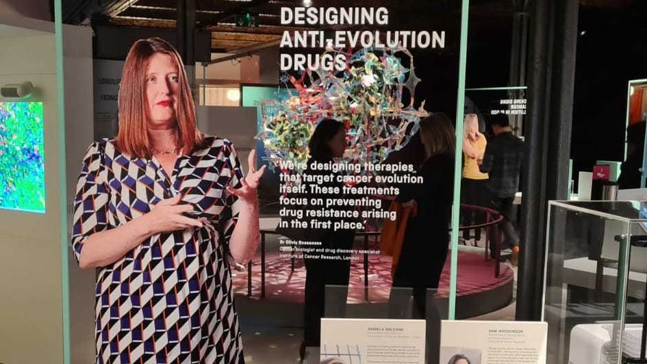
An exhibit in the Cancer Revolution exhibition displaying ICR research.

Image: An exhibit featuring Dr Olivia Rossanese, Director of the CRUK Cancer Therapeutics Unit, and her team's research into APOBEC inhibitors.

A new world-first exhibition by the Science Museum Group titled Cancer revolution: science, innovation and hope features a series of pioneering research projects being carried out at The Institute of Cancer Research, London.

The temporary exhibition is the first of its scope, scale and ambition anywhere in the world, and uses ICR research to help it tell a story of how scientific revolution is transforming cancer care. 

It opens at the Science and Industry Museum in Manchester this week, and will move to the Science Museum in London in summer 2022.

Included in the exhibition are extraordinary objects that reveal monumental scientific discoveries made throughout the history of cancer research, as well as current research that aims to transform the future of cancer treatment.

The exhibition features a range of ICR research, from breakthroughs of the last few decades that have already transformed cancer treatment, to the cutting-edge research of today seeking to understand and overcome cancer evolution.

Tackling cancer's ability to evolve

The exhibition showcases an approach researchers are calling ‘evolutionary herding’.

ICR researchers have shown that it is possible to use artificial intelligence (AI) and advanced maths to forecast how cancers will respond when treated with a particular drug. 

By selecting an initial drug treatment, they have found they can force cancer cells to adapt in a way that makes them highly susceptible to a second drug or pushes them into an evolutionary dead end.

Herding cancer cells in this way through sequential use of cancer drugs could either eradicate the disease or turn incurable cancer into a manageable chronic condition.

Featured alongside this research is the pioneering work of ICR scientists who are creating what researchers believe is the world’s first family of drugs to specifically target cancer’s ability to evolve and become resistant to treatment.

These potential drugs are being designed to stop the action of a molecule called APOBEC to reduce the rate of mutation in cancer cells, slow down evolution and delay resistance.

APOBEC protein molecules are crucial to the ability of the immune system to adapt to different infectious diseases – but are also hijacked in over half of cancer types to speed up evolution of drug resistance.

Researchers hope that a new class of APOBEC inhibitors could be given alongside a targeted cancer treatment to ensure it can keep cancer at bay for much longer.

Engineering viruses to kill cancer

ICR researchers are working to harness the body's immune system to kill cancer, using viral immunotherapy – work that also features in the new exhibition.

Talimogene laherparepvec (T-VEC), a modified form of herpes simplex virus type-1, multiplies inside cancer cells and bursts them from within. It has been genetically engineered to produce a molecule called GM-CSF, which stimulates the immune system to attack and destroy the tumour.

T-VEC was the first of a new wave of virus-based drugs to show benefit to patients in a major randomised, controlled phase III trial.

Targeting ovarian and breast cancer

The Science Museum Group also chose to showcase the ICR's innovative research underpinning the development of targeted drug olaparib, which is now transforming the lives of patients with ovarian, breast and prostate cancer.

Olaparib's origins lie in ICR research to understand the genetic causes of inherited breast in the 1990s, when our scientists tracked down the BRCA2 gene.

A decade after the identification of BRCA2, ICR researchers found that targeting a DNA repair protein called PARP was a potential way to kill cancer cells with a faulty BRCA gene. This helped lead to the development of olaparib and other PARP inhibitor drugs.

Transforming patients’ lives

Professor Paul Workman, Professor of Pharmacology and Therapeutics at the ICR, who was a member of the exhibition's advisory board and attended the launch, said:

“I’m delighted to see the ICR’s research showcased in this new, world-first exhibition. The fact that the Science and Industry Museum have chosen to highlight so much of the ICR’s research is a testament to how our discoveries have and continue to transform the lives of people with cancer. It’s been a real pleasure to be part of it.”

Cancer revolution: science, innovation and hope is open at the Science and Industry Museum in Manchester until March 2022. It will open at the Science Museum in London in May 2022.