Imaging for Radiotherapy Adaptation Group

Dr Emma Harris’ group is investigating ways to make radiotherapy more patient-specific.

Our group develops new image processing techniques and new implementations of ultrasound technologies to image soft tissues and to estimate their motion during therapy.

Dr Emma Harris and her group are developing imaging techniques to increase the effectiveness of radiation oncology to treat a number of different cancer sites. Her main focus is the development of various novel ultrasonic imaging techniques to locate the radiotherapy target volume, to guide the delivery of radiotherapy and to help predict, and monitor, treatment response.

Quantification of Radiation-Induced Tissue Fibrosis using Ultrasound Imaging

Radiation-induced fibrosis is a chronic side effect of radiotherapy given to patients with cancer and may limit the dose that is given. Fibrosis is a genetically regulated response to tissue injury and drugs may be given to reduce its severity. Reliable measures of fibrosis are required to support research in three specific areas: i) radiotherapy dose scheduling, ii) identification of patients’ genetic susceptibility to fibrosis, and iii) clinical evaluation of anti-fibrotic therapies. Quantitative in vivo measures of treatment response are needed to support the investigation of genetic and physiological susceptibility to radiation toxicity and clinical trials evaluating anti-fibrotic, anti-angiogenic and anti-hypoxia therapies.

This research project investigates the potential of new ultrasound based tools in vivo for assessment of radiation induced fibrosis in women following radiotherapy for breast cancer.

<button>Read more<button>

Development of ultrasound motion estimation techniques to optimise radiation therapy delivery

Dr Tuathan O’Shea is researching novel ways to use imaging techniques to improve radiation therapy delivery. It is well known that for many anatomical sites the tumour remains mobile during radiation therapy (RT) requiring margins around the target to account for motion (known as intra-fraction motion). With the application of imaging and improved information on tumour position, margins can be decreased, reducing radiation to surrounding healthy tissue and potentially allowing increased tumour dosage.

<button>Read more<button>

Development of ultrasound guided radiotherapy of the cervix and kidneys

Highly conformal radiotherapy planning techniques such as IMRT and VMAT are only possible if the precise positions of the target volume and organs at risk are known. However, the motile and deformable characteristics of soft tissue organs make it extremely difficult to ascertain their position (despite daily x-ray imaging and careful patient set up) prior to and during radiation delivery. Therefore, large treatment margins are required to ensure adequate coverage of the target area at the expense of including healthy tissues in the treatment field. Daily soft-tissue imaging using ultrasound is a promising solution for determining the precise location of the target organ and surrounding tissues for radiotherapy, which could ultimately allow a more widespread use of highly conformal delivery plans.

<button>Read more<button>

Multi-parametric ultrasound imaging for assessment of tumour response to radiotherapy

Radiotherapy is an important treatment for cancer with around 50% of cancer patients receiving radiotherapy. Unfortunately, not all tumours respond in the same way. Some tumours may be radiation resistant resulting in treatment failure. It is important that we find accurate and cost effective methods to monitor the response of tumours to radiotherapy at multiple times throughout treatment. That way, the clinician can know if the treatment is failing and this allows them to change the patient’s treatment accordingly and quickly. Functional ultrasound imaging techniques such as contrast enhanced ultrasound, molecular ultrasound and elastography, show great promise for the evaluation of tumour response to therapy. They are also affordable and quick to perform making. This project will develop 3D ultrasound imaging which can measure multiple ultrasound characteristics of tumours which may be used to evaluate tumour response to therapy.

<button>Read more<button>

3D ultrasound contrast imaging biomarkers for assessment of tumour response to cancer therapies

Ultrasound contrast imaging is an effective method for monitoring the response of the tumour vascular system to cancer therapies such as targeted drug therapies which target the tumour blood supply, radiotherapy and chemotherapy. One limitation of the techniques is that it is a 2D imaging technique, typically allowing only a small portion of the tumour to be evaluated. We are developing 3D ultrasound contrast imaging which will allow us to evaluate the response of the entire tumour and to look for heterogeneity in tumour response, which is believed to contribute to treatment resistance.

<button>Read more <button>

Radiation treatments are planned using Computed Tomography (CT) scans of the patients. These images allow us to plan the radiation beam arrangement to target the cancer and avoid other tissues, based on the position of the anatomy at CT scanning. Changes in anatomy between CT scanning and treatment, and motion of tissues during radiation delivery means that the treatment may not be delivered exactly as planned. These changes decrease the accuracy of radiotherapy and increase the likelihood, and in some cases, the severity of treatment toxicity. By imaging the radiotherapy target, just prior to and during the treatment, we can ensure that the radiation is safely delivered as planned. Furthermore, some cancers will respond to radiotherapy and some cancers may not respond. Clinicians would like to be able to understand, as soon as possible which tumours are responding and which are not, allowing them to alter the treatment for the best chance of success, or to stop the treatment if the patient will not benefit.

Dr Harris’s group develops new image processing techniques, new implementations of established, and novel, ultrasound technologies to image soft tissues and to estimate their motion during therapy. Ultrasonic elastography and ultrasound backscatter characterisation techniques are being developed for the purpose of assessing the response of cancer and normal tissues to treatments of breast and head and neck cancers. The group, are exploring further integration of functional and molecular ultrasound imaging into the radiation treatment room techniques. Multi-parametric ultrasound imaging has potential to be an affordable and easily implantable method of monitoring tumour response to radiotherapy.

In collaboration with clinical colleagues at the Royal Marsden Hospital the group are currently testing a number of these techniques in clinical trials. These techniques enable more accurate delivery of radiation therapy that spares normal tissue from radiation damage and helps improve the efficacy of radiation therapy.

Dr Harris is a state registered clinical scientist and is a member of the NCRI Breast Clinical Studies Group.

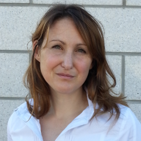
Dr Emma Harris

Group Leader:

Imaging for Radiotherapy Adaptation Emma Harris

Dr Emma Harris and her group are developing imaging techniques to increase the effectiveness of radiation oncology to treat a number of different cancer sites. Her main focus is the development of various novel ultrasonic imaging techniques to locate the radiotherapy target volume, to guide the delivery of radiotherapy and to help predict, and monitor, treatment response.

Researchers in this group

.

Phone: +44 20 3437 6206

Email: [email protected]

Location: Sutton

.

Email: [email protected]

Location: Sutton

.

Phone: +44 20 3437 6328

Email: [email protected]

Location: Sutton

.

Email: [email protected]

Location: Sutton

Dr Emma Harris's group have written 83 publications

Most recent new publication 9/2024

See all their publications

Industrial partnership opportunities with this group

Opportunity: Novel non-invasive imaging tool for measuring tumour stiffness

Commissioner: Dr Emma Harris

Recent discoveries from this group

28/06/16

An ultrasound examination (photo: Jan Chlebik/the ICR)

Puzzling over bats flying at night, Lazzaro Spallanzani — an Italian biologist and physiologist — was one of the first to investigate how ultrasound worked back in the 1790s.

Blindfolding the bats did not stop them navigating through the night, but when their ears were plugged they started bumping into obstacles. Spallanzani concluded that their primary mode of navigation was mediated through waves that echoed off of objects and helped to determine their distance.

Fast-forward around 200 years and we are now using ultrasound in an array of cutting-edge medical applications, including finding, diagnosing and treating cancer.

New ultrasonic imaging techniques

Here at The Institute of Cancer Research, London, Dr Emma Harris and the Imaging for Radiotherapy Adaptation Team are developing various novel ultrasonic imaging techniques to help plan and guide the delivery of radiotherapy and to predict and monitor treatment response.

One of Dr Harris’ projects is using ultrasound to assess changes in breast tissue linked to radiotherapy. Because more women are now surviving breast cancer due to improvements in treatments, late side-effects from radiotherapy are becoming more apparent.

Around 20% of women who receive breast radiotherapy will experience breast hardening or shrinkage. Researchers think that the likelihood of developing these reactions is down to the individual genetic make-up of patients.

emma-harris-feature infographic midarticle

But in order to identify these genetic differences, we need to quantify accurately the severity of these side-effects.

Dr Harris says: “So far, clinicians have judged the severity of the symptoms through touch and visual appearance. But these clinical methods are subjective and can vary greatly. For example, clinical assessment often fails to distinguish between swelling — an early transient effect — and hardness, which develops later and is usually permanent.

“Because of this, we have difficulty in pooling data from different studies, which is an essential approach for investigating genetic susceptibility.”

Ultrasound techniques, however, may be able to help quantify and characterise radiotherapy-associated changes to a higher specificity in breast tissue, allowing researchers to link these with genetic abnormalities.

Pilot studies

Collaborating with Dr Jeff Bamber at the ICR, Dr Anna Kirby’s team at The Royal Marsden NHS Foundation Trust, and clinicians in Addenbrookes Hospital in Cambridge, Dr Harris is conducting the QuaRTUS triala pilot study that’s investigating whether an ultrasound-based imaging technique can measure tissue stiffness accurately.

Researchers are using two approaches: ultrasound backscatter spectroscopy and shear-wave elastography.

Ultrasound backscatter spectroscopy uses the frequencies of ultrasound reflected by tissues to characterise tissue structure, and has already been tested in a breast tissue. Although the technique was able to detect some damage, measurements were difficult to obtain because of variation in ultrasound signal due to differences in breast tissues.


Variation in tissue stiffness midarticle

Breast ultrasound image from a healthy volunteer with elastography information overlaid in colour acquired using Aixplorer – a system that incorporates shear wave sound speed to measure stiffness in breasts. Stiffness is represented by the colour spectrum ranging from dark red (very stiff) through to blue (very soft), and shows higher stiffness in the skin region and lower stiffness in subcutaneous fat. Grey scale image shows a mixed echo texture due to varying tissue architecture in the breast (image: Dr Emma Harris).

Using ultrasound shear-wave elastography, which measures tissue stiffness and stretchiness, does not suffer these problems and may be a preferable alternative.

Dr Harris explains: “Patients who have swelling or hardening of breast tissue after radiotherapy will be tested using the two techniques. We believe that the ultrasound shear-wave elastography will be able to distinguish between swelling and hardness, and will help us determine side-effects from radiotherapy more accurately than ultrasound backscatter spectroscopy. We are eagerly looking forward to the results which are expected in another two years.

“Breast hardening or shrinkage has a heavy influence on a woman’s quality of life after breast therapy,” says Dr Harris. “Being able to recognise and alleviate these symptoms will make a huge difference to patients.”

Tracking radiotherapy

Beyond the QuaRTUS trial, Dr Harris is also addressing the problem of motion and organ movement when delivering radiotherapy. X-rays can be used to guide radiotherapy planning, but these are hard to take during treatment, often not showing soft tissue clearly, and also give the patient an extra dose of radiation.

By using 4D ultrasound to track radiotherapy, images can be taken in real time, and soft tissues can be visualised clearly while putting the patient at no extra risk. “We can process internal tissue movement in real time, so can adjust the radiotherapy accordingly,” explains Dr Harris. “We are now testing this in prostate and cervical cancer studies, but hope to include liver and paediatric cancer patients in the future.”

Much like bats navigating in the dark, ultrasound is now being used to help navigate the path of cancer treatment. However, this is just one side of ultrasound’s many facets — we are also using it to locate tumours and alleviate bone cancer pain — making it a growing and formidable force in cancer research.