Imaging for Radiotherapy Adaptation Group

Dr Emma Harris’ group is investigating ways to make radiotherapy more patient-specific.

Our group develops new image processing techniques and new implementations of ultrasound technologies to image soft tissues and to estimate their motion during therapy.

Dr Emma Harris and her group are developing imaging techniques to increase the effectiveness of radiation oncology to treat a number of different cancer sites. Her main focus is the development of various novel ultrasonic imaging techniques to locate the radiotherapy target volume, to guide the delivery of radiotherapy and to help predict, and monitor, treatment response.

Quantification of Radiation-Induced Tissue Fibrosis using Ultrasound Imaging

Radiation-induced fibrosis is a chronic side effect of radiotherapy given to patients with cancer and may limit the dose that is given. Fibrosis is a genetically regulated response to tissue injury and drugs may be given to reduce its severity. Reliable measures of fibrosis are required to support research in three specific areas: i) radiotherapy dose scheduling, ii) identification of patients’ genetic susceptibility to fibrosis, and iii) clinical evaluation of anti-fibrotic therapies. Quantitative in vivo measures of treatment response are needed to support the investigation of genetic and physiological susceptibility to radiation toxicity and clinical trials evaluating anti-fibrotic, anti-angiogenic and anti-hypoxia therapies.

This research project investigates the potential of new ultrasound based tools in vivo for assessment of radiation induced fibrosis in women following radiotherapy for breast cancer.

<button>Read more<button>

Development of ultrasound motion estimation techniques to optimise radiation therapy delivery

Dr Tuathan O’Shea is researching novel ways to use imaging techniques to improve radiation therapy delivery. It is well known that for many anatomical sites the tumour remains mobile during radiation therapy (RT) requiring margins around the target to account for motion (known as intra-fraction motion). With the application of imaging and improved information on tumour position, margins can be decreased, reducing radiation to surrounding healthy tissue and potentially allowing increased tumour dosage.

<button>Read more<button>

Development of ultrasound guided radiotherapy of the cervix and kidneys

Highly conformal radiotherapy planning techniques such as IMRT and VMAT are only possible if the precise positions of the target volume and organs at risk are known. However, the motile and deformable characteristics of soft tissue organs make it extremely difficult to ascertain their position (despite daily x-ray imaging and careful patient set up) prior to and during radiation delivery. Therefore, large treatment margins are required to ensure adequate coverage of the target area at the expense of including healthy tissues in the treatment field. Daily soft-tissue imaging using ultrasound is a promising solution for determining the precise location of the target organ and surrounding tissues for radiotherapy, which could ultimately allow a more widespread use of highly conformal delivery plans.

<button>Read more<button>

Multi-parametric ultrasound imaging for assessment of tumour response to radiotherapy

Radiotherapy is an important treatment for cancer with around 50% of cancer patients receiving radiotherapy. Unfortunately, not all tumours respond in the same way. Some tumours may be radiation resistant resulting in treatment failure. It is important that we find accurate and cost effective methods to monitor the response of tumours to radiotherapy at multiple times throughout treatment. That way, the clinician can know if the treatment is failing and this allows them to change the patient’s treatment accordingly and quickly. Functional ultrasound imaging techniques such as contrast enhanced ultrasound, molecular ultrasound and elastography, show great promise for the evaluation of tumour response to therapy. They are also affordable and quick to perform making. This project will develop 3D ultrasound imaging which can measure multiple ultrasound characteristics of tumours which may be used to evaluate tumour response to therapy.

<button>Read more<button>

3D ultrasound contrast imaging biomarkers for assessment of tumour response to cancer therapies

Ultrasound contrast imaging is an effective method for monitoring the response of the tumour vascular system to cancer therapies such as targeted drug therapies which target the tumour blood supply, radiotherapy and chemotherapy. One limitation of the techniques is that it is a 2D imaging technique, typically allowing only a small portion of the tumour to be evaluated. We are developing 3D ultrasound contrast imaging which will allow us to evaluate the response of the entire tumour and to look for heterogeneity in tumour response, which is believed to contribute to treatment resistance.

<button>Read more <button>

Radiation treatments are planned using Computed Tomography (CT) scans of the patients. These images allow us to plan the radiation beam arrangement to target the cancer and avoid other tissues, based on the position of the anatomy at CT scanning. Changes in anatomy between CT scanning and treatment, and motion of tissues during radiation delivery means that the treatment may not be delivered exactly as planned. These changes decrease the accuracy of radiotherapy and increase the likelihood, and in some cases, the severity of treatment toxicity. By imaging the radiotherapy target, just prior to and during the treatment, we can ensure that the radiation is safely delivered as planned. Furthermore, some cancers will respond to radiotherapy and some cancers may not respond. Clinicians would like to be able to understand, as soon as possible which tumours are responding and which are not, allowing them to alter the treatment for the best chance of success, or to stop the treatment if the patient will not benefit.

Dr Harris’s group develops new image processing techniques, new implementations of established, and novel, ultrasound technologies to image soft tissues and to estimate their motion during therapy. Ultrasonic elastography and ultrasound backscatter characterisation techniques are being developed for the purpose of assessing the response of cancer and normal tissues to treatments of breast and head and neck cancers. The group, are exploring further integration of functional and molecular ultrasound imaging into the radiation treatment room techniques. Multi-parametric ultrasound imaging has potential to be an affordable and easily implantable method of monitoring tumour response to radiotherapy.

In collaboration with clinical colleagues at the Royal Marsden Hospital the group are currently testing a number of these techniques in clinical trials. These techniques enable more accurate delivery of radiation therapy that spares normal tissue from radiation damage and helps improve the efficacy of radiation therapy.

Dr Harris is a state registered clinical scientist and is a member of the NCRI Breast Clinical Studies Group.

Dr Emma Harris

Group Leader:

Imaging for Radiotherapy Adaptation Emma Harris

Dr Emma Harris and her group are developing imaging techniques to increase the effectiveness of radiation oncology to treat a number of different cancer sites. Her main focus is the development of various novel ultrasonic imaging techniques to locate the radiotherapy target volume, to guide the delivery of radiotherapy and to help predict, and monitor, treatment response.

Researchers in this group

.

Phone: +44 20 3437 6206

Email: [email protected]

Location: Sutton

.

Email: [email protected]

Location: Sutton

.

Phone: +44 20 3437 6328

Email: [email protected]

Location: Sutton

.

Email: [email protected]

Location: Sutton

Dr Emma Harris's group have written 83 publications

Most recent new publication 9/2024

See all their publications

Industrial partnership opportunities with this group

Opportunity: Novel non-invasive imaging tool for measuring tumour stiffness

Commissioner: Dr Emma Harris

Recent discoveries from this group

02/06/20

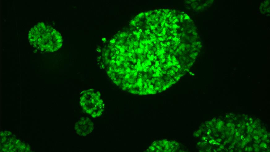
Mini tumours grown from a bowel cancer

Mini tumours grown from a bowel cancer

Scientists have shown that therapies which combine heat and radiation have a different effect on lab-grown mini tumours than they do on lab-grown cancer cells grown in a more traditional way in a two-dimensional layer. 

In a new study, scientists at The Institute of Cancer Research, London, tested the effects of a combination of hyperthermia and radiation on mini tumours grown in what’s called a spheroid – where cells are packed in a 3D structure, much like a small ball.

Cells at the centre of the ball have less access to oxygen and nutrients compared to those on the outside, and this closely mimics tumours in the real world. 

Making cells more vulnerable to treatments

Hyperthermia involves exposing cells to high temperatures, for example through high-intensity therapeutic ultrasound (HIFU) – a pioneering technology developed at the ICR. 

Scientists are exploring the potential of combining hyperthermia and radiation because heating could make cells become more vulnerable to standard radiation treatments given for cancer. 

It is thought that the application of heat increases the blood circulation to the cancer cells, thereby increasing the amount of oxygen available to them, which in turn could make the cells easier to kill with radiation.

ICR is a pioneer in use of mini-tumours

In a series of experiments, the researchers exposed mini tumours to 47 degrees C, radiation, and a combination of both. They repeated these treatments for standard cancer cells grown in dishes in the lab to allow them to compare the effects of the treatments between the two.

They found that cells in the mini tumours are more resistant to heat than standard lab-grown cancer cells. It was also found that in some cases, following heat treatment, cells in mini tumours were inclined to grow at a faster rate. 

This may be because changes in the environment around the tumour made more oxygen available to the centre of the mini tumour, leading to increased growth. 

Based on these results, the scientists believe more work is needed to calculate what the best ‘fractions’, or doses, of radiotherapy given to patients in combination with heat treatment could be. This work should be more closely based on data from experiments with mini tumours rather than that from layers of cells. 

The ICR has pioneered the use of mini tumours, now in use in many labs, which more closely model the growth of a real tumour than two-dimensional cell cultures and also help limit the use of animals in research.

The ICR has an outstanding record of scientific achievement dating back more than 100 years. Find out more about some of our most influential discoveries, all of which have played an important role in shaping our modern understanding of cancer and how it can be treated.

Find out more

’We have been at the forefront of developing this treatment path’

Study co-leader Professor Gail ter Haar, Professor of Therapeutic Ultrasound at the ICR, said: 

“The main aim of our study was to assess the response of tumour spheroids to hyperthermia, radiation, and a combination of the two treatments, and to see if the results were able to give us an accurate picture of how sensitive the cancer cells are to radiation after heat treatment. 

“Heat-based treatments are effective at rapidly killing tissues when delivered via high intensity therapeutic ultrasound, and we have been at the forefront of developing this treatment path. 

“In this study we were looking specifically at the ability of heat to make cells more susceptible to treatment with radiation, and it looks like further work is needed to improve this in the clinic.”

Study co-leader Professor Uwe Oelfke, Deputy Head of Division and Head of the Joint Department of Physics, said: 

“Adding rapid heating of tumours to radiotherapy could make it more effective in some circumstances, through making cells more vulnerable to radiation treatment.

“This study focused on designing new tumour spheroids that could help us gain a much better insight into the effects of heating on cancer cells, and showed that our new models give a more accurate picture of how radiation and heating really affects tumours. Further experiments will give us a better understanding of  exactly which combination of treatments might work.”