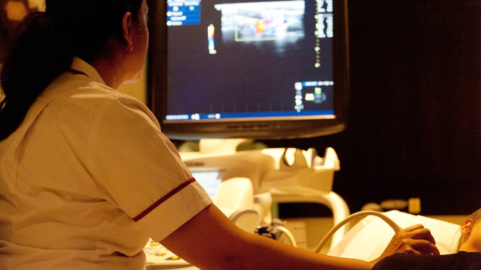
Image: Ultrasound is used to image regions of the body, and could help guide radiotherapy treatment. Credit Jan Chlebik, The ICR
A computer algorithm which helps radiographers interpret ultrasound images could improve radiotherapy treatment for patients with prostate cancer, a new study suggests.
The new algorithm helps radiographers – who are experts in interpreting scans – to quickly locate the prostate in ultrasound scans, which can be time consuming to interpret on their own.
Importantly the algorithm, described in a new study led by scientists at The Institute of Cancer Research and our partner hospital The Royal Marsden and published in the journal Radiotherapy and Oncology, could help radiographers less familiar with ultrasound use the technology to deliver more accurate radiotherapy for prostate cancer.
A safe, effective way to improve radiotherapy
New radiotherapy techniques aim to deliver large doses of radiation in fewer sessions overall, which can be more effective and convenient for patients than smaller doses delivered more often.
Larger radiation doses kill more cancer cells and mean patients need fewer visits to hospital, but treatment also needs to be as accurate as possible to limit damage to healthy tissue.
Radiotherapy is usually guided by X-ray images taken during a CT scan or CAT scan, which are used to build 3D images of metallic markers that are implanted into the prostate.
Ultrasound, which does not require markers, could be as effective, and because ultrasound is safer than X-rays, it has the advantage that it can be used throughout treatment, ensuring the prostate stays in the correct position throughout treatment.
The algorithm helps computer software to compare and match ultrasound scans taken at the planning stage and during treatment, so radiographers could reposition the patient to account for the movement of the prostate inside the body.
In the study, which was funded by Cancer Research UK, the algorithm was used retrospectively on ultrasound scans for 32 patients with prostate cancer treated at The Royal Marsden.
Three technicians then used the scans to select the regions around the prostate to target with radiotherapy.
The algorithm reduced differences in the regions selected by the different technicians and increased the accuracy of radiotherapy targeting, and reduced the time required.
We've lost many vital research hours to the coronavirus crisis but the need for our work continues to grow. Please help us kick-start our research to make up for lost time in discovering smarter, kinder and more effective cancer treatments, and to ensure cancer patients don't get left behind.
Paving the way for more widespread ultrasound use to guide radiotherapy
In the future, the team hopes to develop machine learning techniques to help radiographers who are unfamiliar with ultrasound, enabling them to more easily acquire high quality images and interpret them in real-time. This could make ultrasound a safe, easy and reliable way to guide radiotherapy treatment for the prostate cancer.
Study leader Dr Emma Harris, Team Leader in Imaging for Radiotherapy Adaptation at The Institute of Cancer Research, said:
“Ultrasound is an affordable and readily available technology for monitoring patient health, but in the radiotherapy setting it’s still relatively uncommon. Radiotherapy practitioners are more familiar with using CT scans to guide treatment and ultrasound produces images of the body that look quite different, so helping users identify tissues accurately and consistently is key to advancing its use.
“Our new algorithm helps radiographers match ultrasound scans, and because ultrasound doesn’t use radiation, you could regularly monitor prostate motion during treatment to update radiotherapy, unlike with currently used CT. Our study could pave the way for more widespread ultrasound use to guide radiotherapy for prostate cancer and other diseases.”

 .
.