Dynamical Cell Systems Group
Professor Chris Bakal’s group uses genomic approaches and computational modelling to understand how complex biochemical signalling networks are ‘rewired’ during the development of cancer.
Our goal is to understand how cancer cells determine their shape. The ability of cancer cells to adopt different shapes is not only absolutely essential for metastasis; but is also important to other key aspects of cancer progression such as uncontrolled proliferation, drug resistance, and the inflammatory and immune responses.
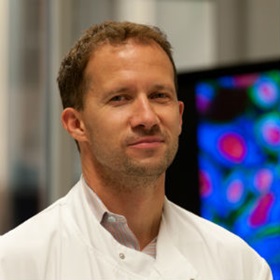
Professor Chris Bakal
Group Leader:
Dynamical Cell Systems
Professor Chris Bakal aims to understand how cells change shape, become cancerous and form metastases. Professor Bakal is a Reader of Cell Form at the ICR and has received numerous awards for his research.
Researchers in this group
Professor Chris Bakal's group have written 82 publications
Most recent new publication 10/2024
See all their publicationsResearch overview
By understanding the systems that determine cell shape, the Bakal laboratory hopes to identify new ways to prevent cancer cells from changing this and open up new therapeutic avenues.
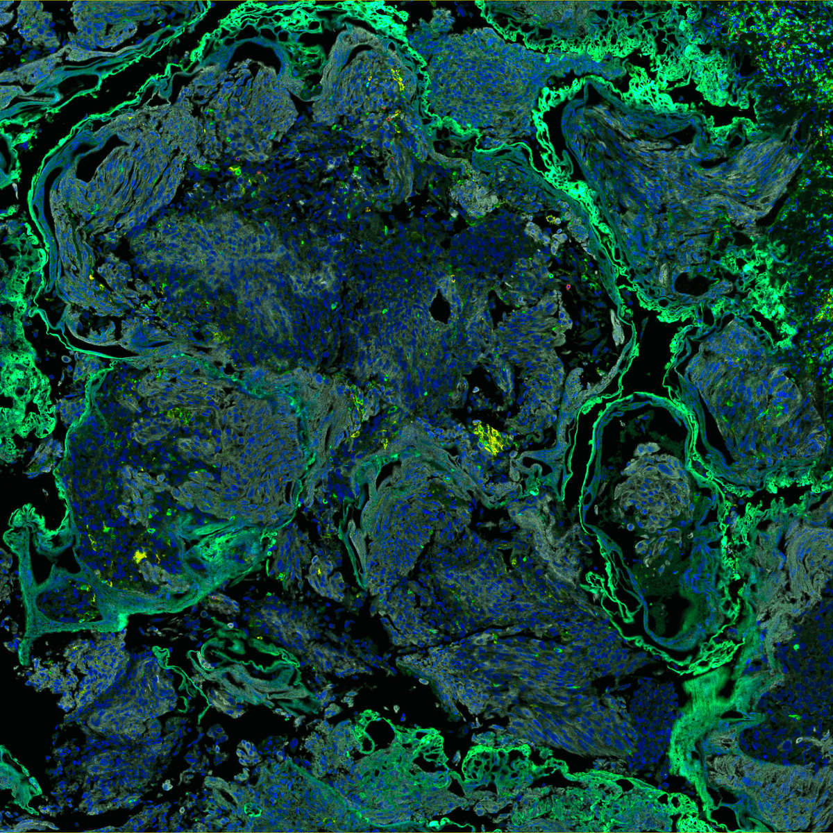
To study cancer cell shape determination the Bakal laboratory uses a combination of cutting-edge microscopy, functional genomic, mathematical, and bioengineering methods. In particular Professor Bakal has pioneered the use of artificial intelligence and new statistical tools to analyse big datasets generated by large-scale imaging.
Currently, work in the Bakal laboratory is focused around the following questions:
1) How do metastatic cancer cells sense, or “feel”, the different complex environments they encounter as they spread throughout the body (i.e. soft versus stiff tissues), and how to they change their shape in response to these different environments in order to metastasize?
Work in this area involves a diverse spectrum imaging approaches - from high-throughput 3D and 4D imaging, to super-resolution microscopy; as well as bioengineering methods to create materials and surfaces that mimic complex environments metastatic cells would encounter in the body.
2) How do biochemical signalling networks determine fate decisions at the single cell level?
The Bakal laboratory uses a number of technologies that monitor and quantify signalling events in single living cells, which are then used to inform statistical and mathematical models which aim to predict, given a set of starting conditions, what cancer cells will do in the future.
3) Can cell shape be used as a diagnostic tool?
By performing integrative analysis of different big datasets, the Bakal laboratory aims to develop technologies that will allow clinicians to diagnose cancer at the single cell level – simply by looking at cell shape.
Research projects
Professor Chris Bakal, Dynamical Cell Systems Group
To describe cellular signaling systems, the Bakal lab uses a combination of functional genomic, proteomic, and computational technologies.
Our research focuses on three main areas:
This research focuses on the networks regulating cell division and mathematical modeling of the cell cycle.
The Networks Regulating Cell Division
Cytokinesis is a key event in the cell cycle requiring tight coordination of mitotic spindle assembly, actomyosin contractility and membrane dynamics, but the mechanisms that regulate cytokinesis are poorly understood. If chromosomes are not accurately segregated during division, and daughter cells lose or gain genomic DNA, this can lead to aneuploidy. Aneuploidy is not only a hallmark of tumor cells but has a causal role in tumorigenesis.
Failures in chromosome segregation can occur if: the mitotic spindle is not properly assembled; cytokinesis is not completed; the spindle assembly checkpoint is inappropriately bypassed; or the timing of late mitotic events is disrupted.
In fact altered expression or mutation of genes that encode proteins regulating the progression of late mitotic events and cytokinesis, such as Aurora-A, Eg5, BubR1, Mad2, or Polo-like kinase (PLK), has been detected in a number of cancers including breast, colorectal, ovarian and lung cancer. Mutations in these genes may override checkpoints that would limit the proliferation of genetically abnormal tumor cells.
Because of the role of aneuploidy in oncogenesis, gaining a systems-levels understanding of the signaling networks that act to ensure the fidelity of chromosome segregation is warranted. Moreover, as a number of anti-mitotic therapies are being devised for cancer therapy it is essential to gain insight in the basis of why these compounds are effective.
In nearly all metazoan cells, the RhoGAP MgcRacGAP and its binding partner the RhoGEF Ect2 are activated in late mitosis by a number of upstream kinases such as Aurora-B, Cdk1, and Plk1 to establish a localized zone of high RhoA GTPase activity at the presumptive cleavage furrow, which in turn promotes the formation of the contractile actomyosin cytokinetic ring and ultimately cytokinesis.
Recruitment of both MgcRacGAP and Ect2 to the presumptive furrow is essential for cytokinesis to occur. Inhibition of the RhoGAP MgcRacGAP or RhoGEF Ect2 results in chromosome segregation defects in a wide variety of organisms.
Late mitotic kinases phosphorylate and activate MgcRacGAP and Ect2 to signal that spindle-chromosome attachment and chromosome segregation has occurred and that cytokinesis can proceed. Interestingly expression of Ect2 has been observed to be upregulated in many human tumors.
Using high-throughput single cell RNAi screens we have previously identified ~40 kinase and phosphatases which are putative regulators of MgcRacGAP and Ect2 during cytokinesis in Drosophila cells, the majority of which have been previously shown to be mutated or have altered expression in human tumor cells.
We hypothesize that these signaling molecules play a role in coordinating spindle assembly and mitotic checkpoint signaling with the initiation of cytokinesis. We will use quantitative multiplexed live cell imaging to determine the role of these kinases and phosphatases in controlling MgcRacGAP and Ect2 localization and activity.
The datasets generated in the course of these studies will then be used in mathematical modeling approaches to describe the dynamics of cytokinesis regulation.
Mathematical Modeling of the Cell Cycle
In order for multicellular organisms to develop and grow, their cells must divide. Even in adults, some cell division still occurs. For example, stem cells divide to replenish shed skin cells, or immune cells divide in response to infection.
This process of cell division must be strictly controlled so that cells only divide when and where new cells are needed. Uncontrolled cell division leads to a mass of cells, or tumor. The genes that regulate cell division are frequently mutated in cancer - which can enhance, diminish or completely change the function of that gene.
Therefore, an understanding of how cell division is normally controlled, and how this process can be corrupted, is of critical importance if we are to understand and combat cancer.
The cell division cycle of human cells is divided into four discrete and consecutive phases known as: G1, S (DNA synthesis), G2 and Mitosis. Arguably, the first transition of the cell cycle – from G1 into S-phase – is the most critical as this represents commitment to a new round of cell division.
A number of proteins have evolved which can stop or start the cell cycle depending on the conditions in- and outside the cell. If, for example, a cell has incurred DNA damage then these proteins will prevent that cell from progressing through the cell cycle.
These regulatory proteins form a complex biochemical network that must be robust to failure, i.e. the system must maintain its function in the face of perturbations. Robustness is a property of many engineered systems, for example the navigation systems of aeroplanes have a number of failsafe mechanisms built-in to ensure continuity of performance if one component fails.
The control networks that control the cell cycle are similarly robust to different types of failure, one example being the loss of function of a regulator protein due to genetic mutation. The robustness of the cell cycle control network, however, comes at a price: the network can also be “rewired” by genetic mutation to drive cell division even when the environment is not suitable – leading to cancer.
This also means that it is difficult to block cancer cell division using different types of drugs, as cells are able to circumvent a block placed on cell division by co-opting and adapting other intact signaling pathways. Despite decades of research, we still do not understand the properties of the control systems that mediate robustness in the cell cycle in normal cells and how these control mechanisms are changed in cancer cells.
Even though we know many of the genes that are important for cell division, we do not understand how they interact in biochemical networks and how these networks are rewired in cancer cells. We aim to quantify the signaling events occurring during progression from G1 into S-phase in human cells.
In collaboration with the laboratory of Michael Yaffe (Massachusetts Institute of Technology, Cambridge USA) we are using this data to computationally model the networks that control G1 progression in order to understand both why the G1 control network is robust to genetic perturbation and how this robustness is hijacked by oncogenic mutations that drive cancer progression.
This research focuses on the control of reactive oxygen species and systems biology of the unfolded protein response.
Systems biology of the Unfolded Protein Response
Humans evolved the systems to manage energy in conditions where resources were largely scarce and demands on energy were very high. However, many humans now live in society where there is an excess of nutrients especially sugars and fats.
Thus the adaptive mechanisms that manage energy and nutrient use have not evolved to cope with such environmental conditions, and coupled with increasingly sedentary lifestyles and increasing lifespan, the failure to adapt to dietary excess leads to metabolic syndromes such as type 2 diabetes.
The Endoplasmic Reticulum (ER) is the principal organelle dedicated to the folding and transport of a diverse range of newly synthesized proteins. Networks of regulatory enzymes monitor cellular conditions such as the levels of available nutrients, and in turn regulate the capacity of the ER. ER capacity must be tightly controlled in order to ensure that protein production can meet demand, or that energy is not wasted in periods of stress.
One critical system that is engaged to increase ER capacity when the ER is overwhelmed is called the Unfolded Protein Response (UPR). The UPR is an allostatic response that acts in several different ways to enhance the protein-folding function of the ER, decrease the level of peptides inputted into the ER, and promote cellular survival.
However, long-term engagement of the UPR likely plays a role in promoting the onset of chronic inflammatory states that can lead to disease. Yet we know very little of the genes involved in turning the UPR on or off. Identifying these genes in the first step in the design of therapies that can be used to modulate UPR activity during the treatment of diseases such as diabetes that are driven by inflammation.
In higher eukaryotes, the UPR consists of at least three different branches triggered by the stress-sensing proteins ATF6∝, PERK, and the highly conserved IRE1∝. Components of the three branches of the UPR are known to engage in cross talk with multiple signaling networks in both physiological and pathological circumstances, such as those which mediate insulin action and regulate autophagy. Signaling cross-talk likely plays a large role in determining the consequences of UPR activation (e.g. survival versus apoptosis).
As activation and/or deregulation of the UPR has been reported to play a relevant role on complex pathological entities such as type 2 diabetes, neurodegeneration, and cancer, understanding UPR on a systems-level is important for both fully understanding these pathologies and achieving successful therapeutic design. For instance, specific types of cancer such as myeloma may be particularly sensitive to intervention of the UPR.
We are gaining systems-level insights in the role of ER stress in disease by performing RNAi screens for genes involved in all three branches of the UPR. We then aim to generate probabilistic models of the dynamic and hierarchical relationships between these genes through computational integration of functional genomic data with orthogonal datasets.
Control of Reactive Oxygen Species
Reactive Oxygen Species (ROS) such as superoxide (O2-), free radicals, and peroxides are natural by-products of oxygen metabolism that also activate signaling networks regulating a spectrum of cellular behaviors including proliferation, differentiation, death, and morphogenesis. While the notion that ROS are causal to ageing has been questioned as of late, high levels of ROS have been clearly been linked to diseases such as diabetes, cancer, as well as neurodegenerative and cardiovascular disorders.
Eukaryotic cells have evolved a complex regulatory system in order to prevent excessive accumulation of ROS during normal metabolic processes, while maintaining the ability to raise ROS levels as needed.
However little is known about the architecture and dynamics of this system. Because the manipulation of ROS levels represents a powerful strategy to prevent a variety of diverse diseases, gaining a comprehensive and quantitative description of the ROS regulatory system is warranted.
In order to understand how ROS levels are genetically controlled, we have performed a genome-wide RNAi screen for regulators of ROS in Drosophila cells and have identified a number of both known and novel genes that are enhancers or suppressors of superoxide levels.
Inhibition of Drosophila dorsal/dl, which encodes an Nf-kB/Rel protein, was isolated as a suppressor of ROS consistent with previous studies in mammalian systems that have demonstrated an antioxidant function for Nf-kB. Interestingly we also isolated cactus/cact, the Drosophila ortholog of the Nf-kB inhibitor IkB as a ROS suppressor. While inhibition and hyperactivation of Nf-kB signaling lead to similar increases in ROS levels, the underlying biochemical mechanisms for the observed increase are likely very different. To begin to understand these differences, we performed two sets of experiments.
First, we profiled genome-wide mRNA expression in Nf-kB and IkB deficient cells in order to determine the transcriptional targets of Nf-kB that may be involved in regulating ROS levels. Second, we performed a series of screens in Drosophila cells for regulators of ROS where we inhibited kinases, phosphatases, and transcription factors (TFs) in sensitized backgrounds in which either Nf-kB or IkB was also inhibited by RNAi.
That many genes isolated in these sensitized screens are also well-characterized regulators of insulin signaling, metabolism, ER stress, and autophagy strongly suggests these cellular processes are normally involved in Nf-kB-mediated ROS production. But these systems-level studies do not provide insight into the biochemical mechanisms by which Nf-kB, Nf-kB genetic interactors, and Nf-kB transcriptional targets are involved in control of ROS.
We are now: (a) performing studies with the aim of understanding how Nf-kB transcriptional dynamics are regulated by the genetic interactors identified by sensitized RNAi screens for regulators of ROS; (b) determining how Nf-kB and its interactors are involved in the control of ROS levels by acting as regulators of ER stress, insulin signaling and autophagy; (c) assembling a comprehensive genome-wide list of ROS regulators that interact with Nf-kB and IkB; and (d) performing computational integration to develop hierarchical and dynamic systems-level models of the Nf-kB signaling networks which regulate ROS levels.
This research looks at how cancer cells adopt different shapes during metastasis and what genes are responsible for metastasis.
How do Cancer Cells Adopt Different Shapes During Metastasis?
The migration of cells is an essential event during organism development and for many functions in the adult organism. For example immune cells have to migrate to sites of infection in order to eradicate bacteria or viruses. Abnormalities of cell migration are also characteristic of disease processes such as inflammatory diseases or metastatic cancers.
In both normal and pathological settings, cells must reorganize their cell shape in highly dynamic and orchestrated manners in order to migrate. Molecules called Rho-family GTPases act as biochemical switches that couple cytoskeletal organization to distinct environmental signals regulate these shape changes. Rho guanine nucleotide exchange factors (RhoGEFs) and Rho GTPase activating proteins (RhoGAPs) are the principal regulators of Rho-family GTPases.
But the specific signals that activate or inhibit RhoGEFs and RhoGAPs, as well which RhoGTPases are their in vivo targets are largely unknown. Moreover, the downstream effectors of Rho signaling which link Rho activity to specific changes in cell shape (e.g. retraction, or the formations of protrusions and lammelipodia) are poorly understood.
We using are high-throughput combinatorial RNAi screening methods where cell shape is used as a phenotypic readout to identify both upstream regulators and downstream effectors of Rho signaling on a systems-level.
Through previously performed genome scale RNAi screens we have determined the contribution of all RhoGEFs, RhoGAPs, RhoGTPases, kinases, phosphatases, and transcription factors to cell shape. Each gene is assigned a Quantitative Morphological Signature (QMS) which a multi-dimensional vector describing the effects of gene inhibition on morphology.
We are now systematically inhibiting all Rho components in combination with all other, as well as with all known kinases, phosphatases, and transcription factors to determine quantitative genetic interactions that underpin cell shape.
These screens can be used to identify both specific and general regulators of individual Rho components and thus provide novel insight as to how Rho-family GTPase activity is tailored resulting in specific phenotypic output in diverse environmental conditions.
Importantly, performing combinatorial screens with quantitative morphology as a readout requires the development of new computational methods to determine epistastic relationships.
In collaboration with the Berger laboratory (Massachusetts Institute of Technology, Cambridge USA) and the Wong laboratory (Methodist Research Institute, Houston USA) we are continuing to develop these methods and have validated their use in preliminary studies.
What Genes are Responsible for Metastasis?
Complex and highly coordinated changes in morphology occur during cancer cell metastasis. For example, metastatic cancer cells of epithelial origin lose cell-cell contacts and apical-basal polarity, engage once-dormant migratory machinery, remodel the extracellular matrix (ECM) and dynamically regulate integrin-based adhesion.
Importantly, while there are common morphological aspects of metastasis, cells may also alternate between distinct modes of migration, such as mesenchymal versus amoeboid. Understanding how signaling networks that control shape are differentially rewired during oncogenesis is critical for developing safe and effective therapeutics.
Given the recent advances in genomic profiling, we have an unprecedented opportunity to describe the genotype of cancer cells. However, a major challenge in the post-genomic era is to understand which genetic alterations are truly drivers of cancer cell phenotypes. For example, it is not clear which transcriptional changes underpin the ability of metastatic cells to migrate and invade secondary tissues.
A number of studies have identified potential “master” regulators of metastatic phenotypes such as Twist, and RhoC, but the list of downstream effectors that lead to cell shape changes remains far from complete.
One well-characterized example of a link between metastatic cell genotype and phenotype is the switch from E-cadherin to N-cadherin expression that results in a loss of cell-cell contacts and mesenchymal morphology in certain epithelial lines. But we hypothesize there are many more unknown genetic changes that are essential for the morphogenesis of metastatic cells.
We are using methods we have previously developed in order to quantify cell shape in parallel with genome-wide microarray and comparative genomic hybridization techniques to determine how specific, quantitative differences in cell morphology in both 2D and 3D are driven by changes in gene expression and copy number variation.
Recent discoveries from this group


 .
.

