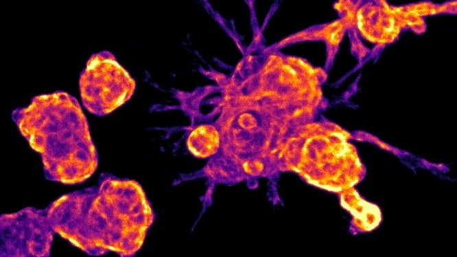
Image: Melanoma cells in their rounded form, taken using stage-scanning oblique plane microscopy. Credit: Professor Chris Bakal
Scientists have discovered how skin cancer cells shapeshift based on their environment – enabling them to spread through the body and cause a metastatic cancer.
The discovery of two genes responsible for sensing the environment and adapting cell shape could offer potential new drug targets to stop cancer from metastasising.
Cancer cells sense where they are
Cancer cells can change shape to move around the body, becoming drill-shaped to ‘poke’ through dense tissue like bone, or round and squishy to squeeze through soft tissues and get into the blood.
In a study published in Cell Reports, scientists at The Institute of Cancer Research, London, have uncovered how cells know what environment they are in and therefore which shape to choose.
Imaging cells in 3D
The team, working in collaboration with researchers at Imperial College London, developed a new system to study cells in 3D – mimicking different parts of the body. Until now, most research has studied cells on hard 2D plastic surfaces.
A unique microscope called stage-scanning oblique plane microscopy (ssOPM) was used to take 3D images of melanoma cells – either stuck to a flat and rigid surface or embedded within a 3D soft collagen hydrogel.
By analysing the 3D images taken of 60,000 cells when certain genes were ‘switched off’, the researchers identified two genes that are important for melanoma cells to change their shape in response to their environment.
Two potential new drug targets to stop cancer spreading
The researchers believe these genes, TIAM2 and FARP1, could be targeted to prevent melanoma cancer from metastasising. These genes are good candidates for drug discovery as they have a structure similar to other proteins for which drugs are already in pre-clinical development.
The research team are currently creating AI-based technologies to make predictions about which drugs might be successful, by using these 3D images of cells – which could cut the time taken to develop a drug in half.
This research was funded by The Institute of Cancer Research (ICR) itself, which is both a research institute and a charity, together with Cancer Research UK, Imperial College London, and The Engineering and Physical Sciences Research Council.
Professor Chris Bakal, Professor of Cancer Morphodynamics at The Institute of Cancer Research, London, said:
“Once cancer becomes metastatic and spreads to different parts of the body, it can be quite difficult to treat. For a metastatic cell, travelling across the body is an epic journey where they encounter many different types of landscapes and environments. Up until now, we haven’t known much about how these cells know which environment they are in, in order to change their shape and continue their journey.
“This research has given us insight into the tricks that cancer cells are using to keep growing and spreading. We’ve identified two genes which could, in the future, be targeted to stop melanoma cancer from changing shape and metastasising.”
Professor Kristian Helin, Chief Executive of The Institute of Cancer Research, London, said:
“By using an innovative technique to study cells as if they were in a human, rather than in a laboratory, our scientists have uncovered a mechanism which cancer cells are using to move around the body. We know that most cancer deaths occur because cancer has spread from the original tumour to other parts of the body. I hope that further research will lead to the development of new treatments for metastatic melanoma.”
Professor Chris Dunsby, Professor of Biomedical Optics at Imperial College London, said:
“This study is the first to apply a new high-content microscopy technique called oblique plane microscopy to study many thousands of cells in 3D. In the future, we hope that the approach demonstrated here can be applied to address a wide range of questions in cancer biology.”