They say a picture paints a thousand words, and the same also holds true in science.
A team led by Dr Chris Bakal, head of the Dynamical Cell Systems Team at The Institute of Cancer Research, London, recently used sophisticated imaging techniques to investigate the Endoplasmic Reticulum (ER), the factory in cells that makes the building blocks they need to grow.
Importantly, this factory has to ‘keep up’ in cancer cells to sustain their uncontrolled growth and increased needs. And a key player controlling these adaptations is SREBP, a protein that orchestrates the metabolism of lipids in many organisms, including flies and humans.

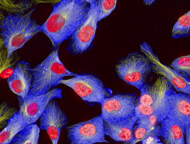
As well as publishing a research paper in the journal PloS One, they produced these striking images of cells, which show how the ER from cells that are increasing their ER capacity physically expands (above), and how this expansion is impaired when SREBP protein is lacking (below).

These images illustrate how SREBP helps regulate the ER, and the BBSRC, who helped fund the study, have posted some of them on their tumblr feed.
And below are more amazing images taken by Dr Bakal’s team of the endoplasmic reticulum in human breast cells:

In the above image of healthy cells, the blue endoplasmic reticulum is clustered around the nucleus, in red. Under normal conditions the ER fills the cells evenly and has a regular structure.

But in these breast cells, the protein SREBP has been silenced, which causes the green endoplasmic reticulum to be much more irregularly shaped. Many of the cells show dense clustering of the ER around the nucleus, while at the edge of the cells, the ER is poorly structured. These are the hallmarks of an ER under stress. The ER of cancer cells are forced to work overtime to help support their out of control growth, so they often display the same features.
These images demonstrate the vital role SREBP plays in controlling how the endoplasmic reticulum operates and the team’s study may help to explain how cancer cells ensure they cover their abnormally high needs, which could one day lead to new treatments for cancer.