Electron Microscopy Facility
The Electron Microscopy Facility in Chelsea brings together a combination of instrumentation and expertise that supports the determination of three-dimensional protein complexes structures by cryo-electron microscopy (cryo-EM).
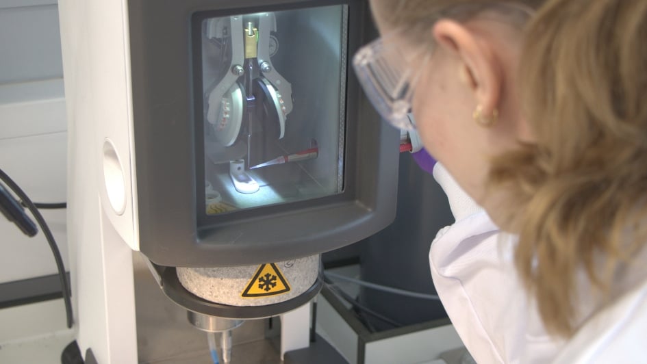
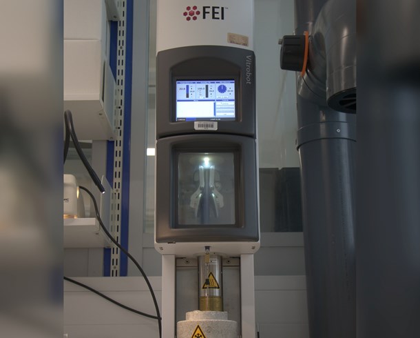
Image: Cryo-EM sample preparation Vitrobot system at the ICR's Electron Microscopy Facility. Credits: ICR
Transmission electron microscopes (TEM)
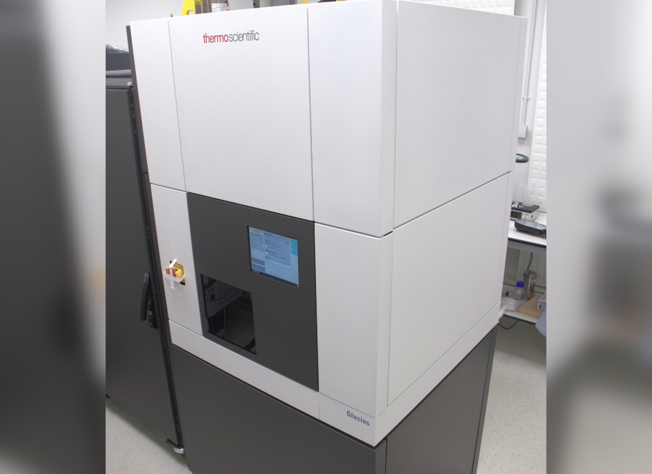
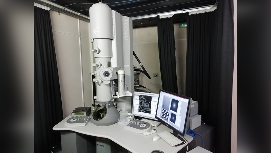
Our Electron Microscopy Facility, based in our labs in Chelsea, houses two ThermoFisher Scientific transmission electron microscopes (TEM) – a Tecnai F20 and a Glacios. It is open primarily to internal users, although also welcomes outside fee-paying users.
The Tecnai F20 microscope is dedicated to the data collection by fully trained users of the Division of Structural Biology both in negative stain and cryo-electron microscopy to moderate resolution.
The state-of-the-art Glacios is dedicated to the data collection of high-resolution data in cryo-electron microscopy and operates at 200 kV. Our Glacios is now equipped with a Falcon 4i direct detector, the first installed in the UK, with higher signal quality and allowing higher throughput.
Cryo-electron microscope (Cryo-EM)
Cryo-electron microscopy has become the dominant technique for protein structure determination in the field of structural biology. The Institute of Cancer Research, London, has long been a pioneer in the field of structural biology and a leading international institution in the use of cryo-EM in cancer research.
Our researchers have access to the ThermoFisher Scientific Titan Krios cryo-EM advanced electron microscope based at the Francis Crick Institute in London, via our membership of LonCEM, the London consortium for cryo-EM – of which the ICR is one of the founding members. Researchers in the ICR’s Division of Structural Biology also have frequent access to the UK national facility at Diamond Light Source (eBIC) through a block allocation group.
In both these facilities the ICR researchers collect data for high-resolution protein structure determination on ThermoFisher Titan Krios 300 kV cryo-electron microscopes.
Focused ion beam scanning electron microscope (FIB-SEM)
The ICR has recently been awarded Biotechnology and Biological Sciences Research Council (BBSRC) ALERT scheme funding for a Focused Ion Beam Scanning Electron Microscope (FIB-SEM). The instrument will be dedicated primarily to state-of-the-art methods for thinning cryogenic biological samples for in situ structural and cellular biology studies by cryo-EM. The £1.5 million award from UKRI, along with contributions from the ICR and our partner institutions Imperial College London, Queen Mary University London, and King’s College London (KCL), will transform our capability to carry out cutting-edge in situ structural biology in central London.
An instrument lab of the ICR’s Electron Microscopy suite is being refurbished for installation of the new FIB-SEM. The FIB-SEM will be capable of thinning lamellae directly in frozen cells and tissue samples; will be capable of lift-out of deep areas of larger frozen samples; and will be capable of destructive volume imaging of ultrastructure by serial FIB-SEM. The FIB-SEM is specified with integrated fluorescence imaging to allow targeting of procedures in samples making use of fluorescence-targeting approaches.
The FIB-SEM completes a pipeline for cutting-edge in situ structural biology studies, encompassing advanced light microscopy infrastructure in ICR’s Light Microscopy Facility, a cross-facility Correlative Light & Electron Microscopy lab with high pressure freezer, cryo-confocal light microscope and cryo-ultramicrotome, and high-resolution cryo-EM for structure determination in ICR’s Electron Microscopy Facility.
Once installed, the FIB-SEM can be accessed by external users subject to project feasibility and resourcing. For more information, please contact Dr Teige Matthews-Palmer.
Sample preparation equipment
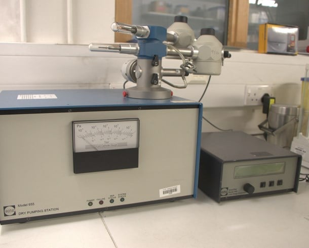
In-house we also have several pieces of equipment for sample preparation, including: Pelco easiglow, Tergeo plasma cleaner, Edwards auto 306 carbon coater, Vitrobot mark IV, Two Gatan 626 cryo-holders, Gatan 655 dry pumping station. We also offer training and advise to students and postdocs in the use of microscopes.
Contact
For more information, please contact our facility manager, Dr Teige Matthews-Palmer.
“Our state-of-the-art Electron Microscopy Facility is a vital component of our research infrastructure and key to training the next generation of Structural Biologists.” - Professor Sebastian Guettler, Team Leader, Structural Biology of Cell Signalling
List of our equipment
The Tecnai TF20 is a 200 kV high resolution TEM, suitable for cryo single particles and semi-thick frozen cells or extract. It enables users to screen samples and obtain preliminary data prior to more detailed analysis on the higher specification Glacios TEM.
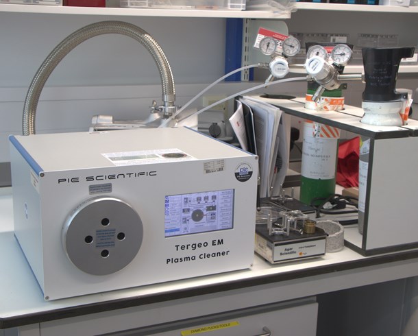
The plasma cleaner uses a mixture of gases to make TEM grid hydrophilic for cryo-EM applications. Unique gentle downstream mode and pulsed plasma can handle ultra-thin carbon and graphene grids without damaging the fragile grids.
Plunge freezing, humidity controlled and automated system (ThermoFisher) for preparation of thin vitreous ice for the structure determination of single particles by cryo-EM.
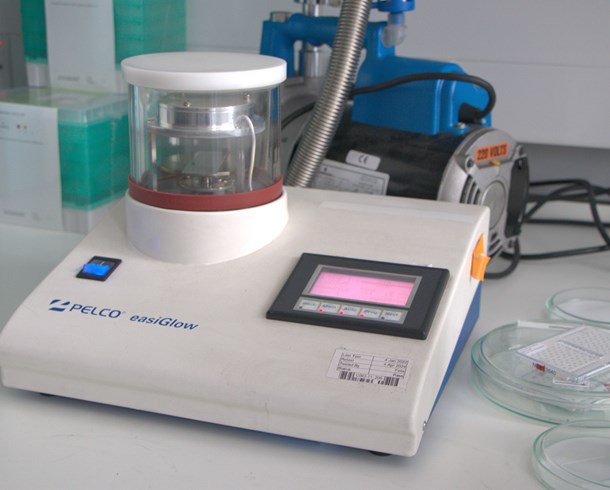
Fully automated system allowing for cleaning and hydrophilisation of carbon support films TEM grids for preparation of TEM grids for negative stain or cryo-EM.
The pumping station is designed to regenerate sorb material in the 626 cryo-holders to allow for the best temperature and stability performance. The 626 single tilt liquid nitrogen cryo-transfer holder is designed for low temperature transfer and subsequent screening and data collection in the Tecnai 12 or TF20.
