Magnetic resonance imaging (MRI) could monitor aggressive cancer that has spread to the bone anywhere in the body, a new study reveals.
Scientists at The Institute of Cancer Research, London, used an MRI technique called quantitative whole-body diffusion-weighted imaging (DWI) to measure skeletal tumours in nine patients with breast and prostate cancer.
They found that the scan identified the spread of cancer in patients’ bones throughout their bodies, with two experts closely agreeing on the number, size and position of tumours.
Quantitative whole-body DWI could monitor how cancer in the bones responds to treatment, which is not possible currently in the clinic.
The study was published in the journal PLOS One and was funded by Cancer Research UK (CRUK) and the Medical Research Council, with additional support from the National Institute for Health Research Biomedical Research Centre at the ICR and The Royal Marsden NHS Foundation Trust.
Using computer algorithms
Secondary bone cancer is one of the biggest killers in cancer. Tumours from diseases like breast and prostate cancer can metastasise to patients’ bones, and it is often difficult for doctors to measure the extent of this spread, or if tumours are responding to treatment.

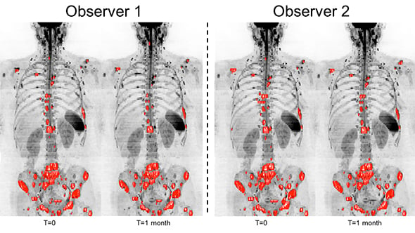
Whole body MRI scans of a breast cancer patient, assessed by two observers, to measure tumours in the patient’s bones. The software could help clinicians reliably assess how bone disease changes over time (image: Blackledge et al 2016)
ICR scientists used quantitative whole-body DWI to monitor bone metastases in nine patients with breast and prostate cancer.
Previously radiologists needed to manually identify tumours inside bones, but the new system uses computer algorithms to help identify cancer more quickly and consistently.
Regions marked as cancer were used to calculate total disease volume for each patient, and this produced consistent results between two experienced observers, and between scans taken one month apart.
'Substantial promise'
Researchers found that a measure called the global apparent diffusion coefficient — which measures water movement in cells and can indicate if tumours respond to treatment — also showed good agreement between observers.
Study leader Professor Martin Leach, Co-Director of the CRUK Cancer Imaging Centre at the ICR, said: “Targeted therapies could prolong the lives of patients with cancer that has metastasised to their skeletons, but it’s challenging to identify which patients gain benefit from these treatments. Our method shows substantial promise for evaluating cancer that has spread to the bone, and for assessing promising new treatments.
“Quantitative whole-body DWI could have a significant impact in drug development and clinical practice, where no reliable method has been established to measure total cancer burden in patients’ skeletons, or to evaluate the response of bone metastases to treatment.”