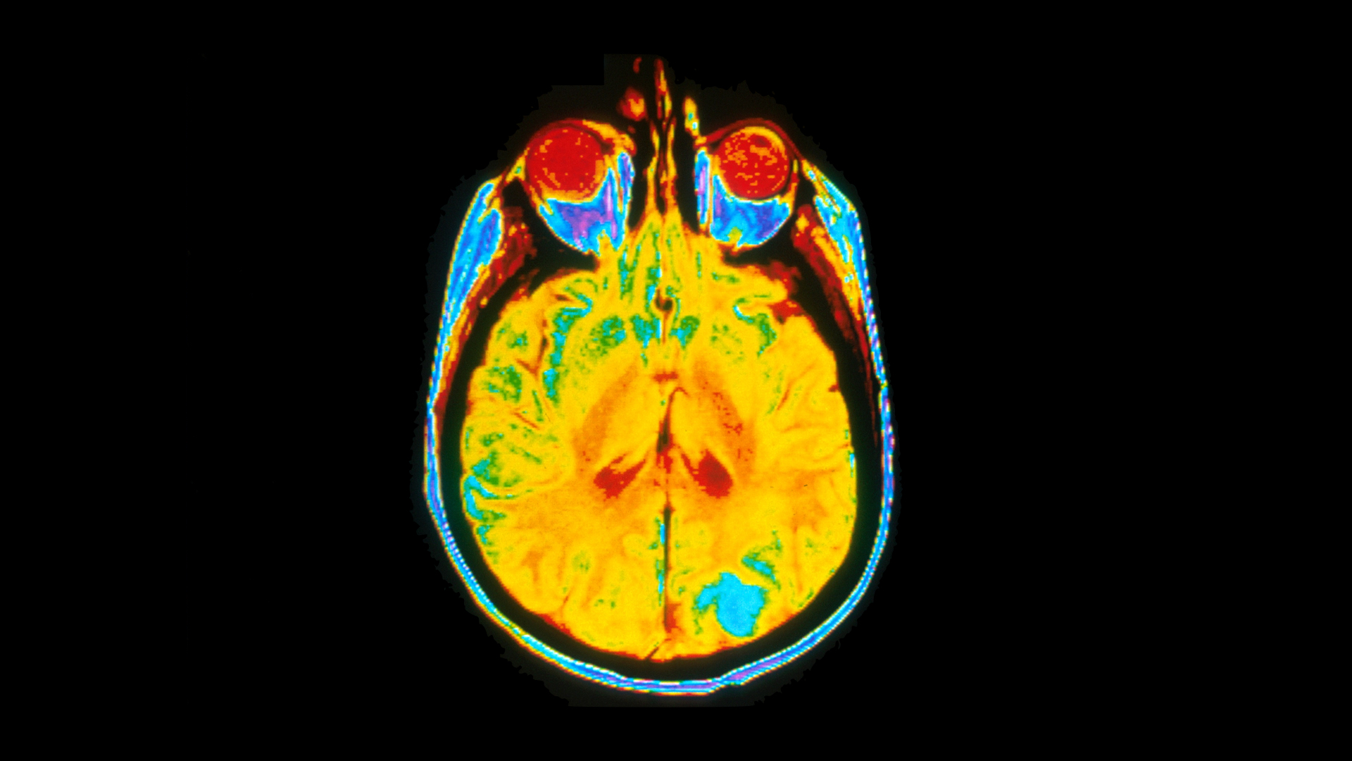
A new type of scan uses a clever ‘noise cancelling’ technique to map cancer metabolism inside brain tumours, a new study shows.
Scientists from The Institute of Cancer Research, London, found the scanning technique could pick up signals for lactic acid – a key indicator of cancer metabolism – while tuning out unwanted noise.
In 20 patients with brain cancer, the scan could clearly distinguish between lactic acid and similar signals produced by molecules of fat, which are found throughout the body.
Lactic acid can build up in muscles during exercise because of a lack of oxygen. Similarly, high levels of lactic acid can also occur in tumours, brought about by low oxygen levels due to their limited blood supply and by higher lactic acid production characteristic of many tumours. Scanning for lactic acid could therefore be a way to detect how tumours respond to treatments targeting cancer metabolism.
The study was published in the journal NMR in Biomedicine, and was funded by Cancer Research UK, with additional support from the Cancer Research UK and EPSRC Cancer Imaging Centre at The Royal Marsden and the ICR.
Standard magnetic resonance imaging (MRI) can map tumour tissue by mapping signals from water molecules in tissues when exposed to a magnetic field.
Other MRI techniques tune in to different molecules, and in cancer, one key molecule to look for is lactic acid, a by-product from a metabolic process called glycolysis which contributes to cancer growth.
But fat molecules produce similar, stronger signals than lactic acid, often drowning it out. Now a new MRI technique called ‘double quantum filtering’ can tune in to lactic acid signals while cancelling out those from fat molecules.
Scientists at the ICR tested the new quantum filtering MRI technique to see if it could accurately measure lactic acid in brain tumours.
They found that the scan was able to suppress 98% of the signal produced by fat molecules in a sample of vegetable oil, while accurately measuring the concentration of lactic acid in a solution of brain metabolites.
They scanned 20 patients with brain tumours using the new technique and they saw a clear lactic acid signal in 15 patients with glioma. They also saw that patients with high-grade glioma had higher levels of lactic acid than the other patients.
The findings show that the new MRI technique could help doctors to identify patients with the most aggressive brain tumours, and could be used to monitor their response to new treatments.
Professor Martin Leach, Leader of the Magnetic Resonance Team at the ICR, said: “Lactic acid is a key product of glycolysis, which cancers harness to fuel their growth despite a limited oxygen supply. A number of promising treatments target lactic acid production in cancer cells, but until now it’s been difficult to monitor their effectiveness because lipids, such as fats and fatty acids that are all around the body, mask their signal.
“What our study found was that this new MRI technique could see lactic acid signals even when lipids were present. In patients with brain tumours we were able to directly scan for lactic acid while simultaneously suppressing the signals from lipids. Our findings demonstrate the effectiveness of this new technique in patients, and show that this could be an important way to assess new cancer treatments that target metabolism.”
