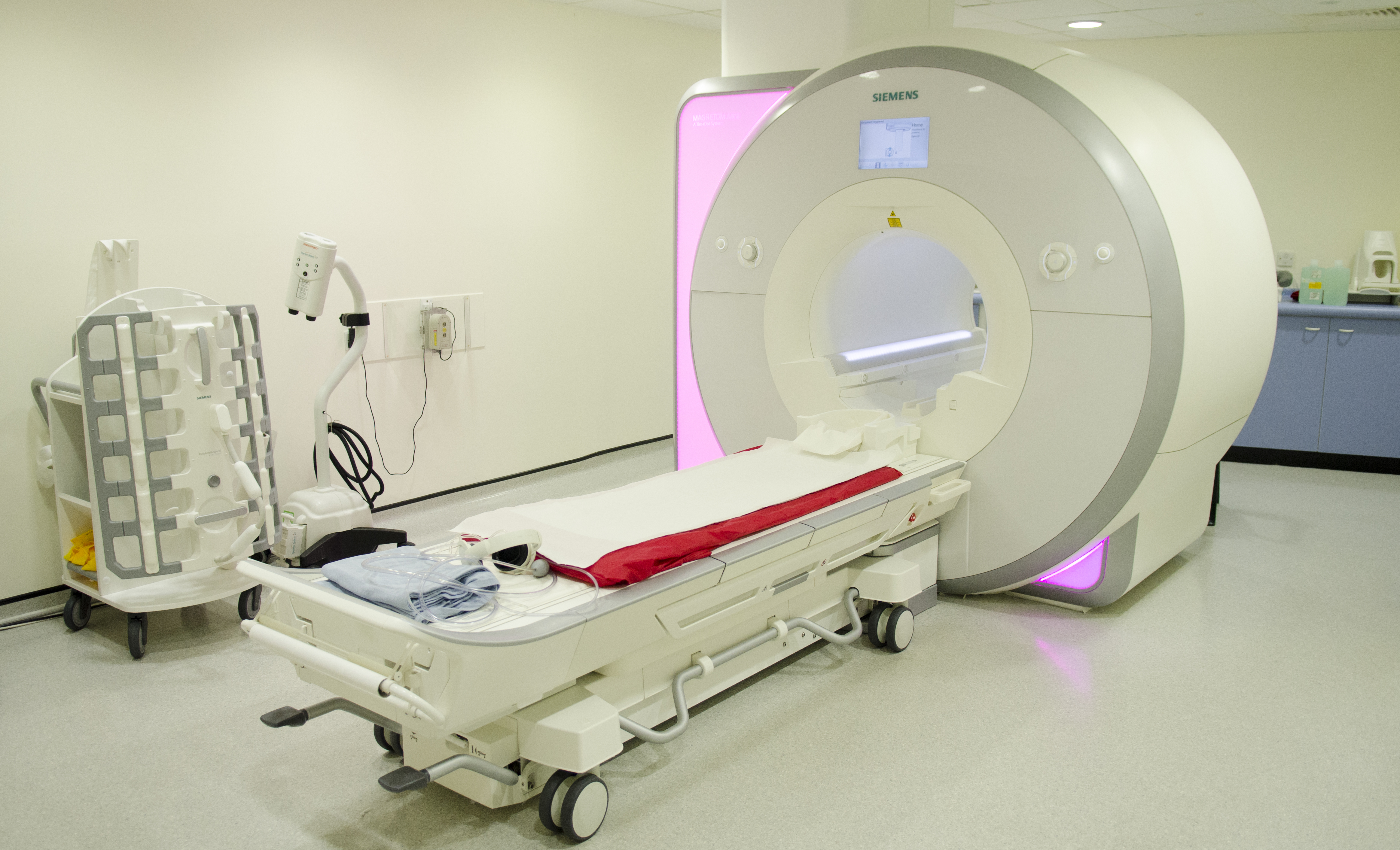
New research suggests an MRI scan could one day help doctors check whether stem cells have transplanted effectively into the brain, say researchers from The Institute of Cancer Research, London.
Implanting neural stem cells (NSC) into the brain of stroke patients is a potential new treatment for repairing brain injury. Researchers found that a type of MRI scan called Proton Magnetic Resonance Spectroscopy (1H-MRS) could successfully track NSCs in cell cultures and was able to show when the cells developed into mature brain cells.
The scientists from the ICR and King’s College London saw that cultures of undifferentiated neural stem cells, the body’s source of new brain cells, displayed a distinct chemical profile to differentiated, or mature, neural stem cells.
They found that the undifferentiated human NSCs had high concentrations of chemicals called phosphophocholine (PC), glycerophosphocholine (GPC) and myo-Inositol (mI), which dropped when the cells became differentiated. Undifferentiated neural stem cells expressed three times more PC and GPC than differentiated cells, and over 50 times more mI. NSCs share many characteristics in their metabolic profile with tumour cells, so 1H-MRS could also be used to evaluate the metabolism of cancer to aid diagnosis and monitor new treatments.
Published in the journal NeuroReport, the study was funded by the Medical Research Council and conducted in the Cancer Research UK and EPSRC Cancer Imaging Centre at The Royal Marsden Hospital and ICR, with additional funding from the NIHR Biomedical Research Centre.
Early stage clinical trials are currently testing whether neural stem cell implants could provide a treatment for patients left with disability after stroke, but improved imaging techniques to monitor the safety and effectiveness of this treatment are needed.
Further research will be required for the technique to be adapted from imaging cells in culture to imaging a live adult brain. However, if the technique is successful, it could provide a non-invasive method to monitor how well NSCs assimilate into the brain and develop into functioning brain cells.
Dr Yuen-Li Chung, senior staff scientist in the Magnetic Resonance team at The Institute of Cancer Research, London, said: “This study shows that 1H-MRS can be used to monitor neural stem cells as they are maturing into functioning neurons. While more research is needed, if the technique can be adapted to the adult brain, it could be used to check how well new treatments for stroke are working.
“The technique also has applications in cancer; different levels of metabolites in tumours are a telltale sign of how the cancer is progressing and how well it is responding to treatment.”
