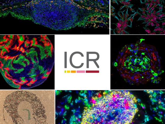
Despite Covid-19, we were thrilled to see a high calibre of entries to our annual imaging competition, which celebrates the incredible research led at the ICR and The Royal Marsden, and the arresting images they can produce.
The selection chosen for the judges’ shortlist cover a range of cancer research fields and various scientific techniques to striking effect – including mouse xenografts, tumour spheroids and immunohistochemistry staining – to tell a story about our science.
The shortlist also features online and in print in New Scientist magazine, which is a testament to the appeal of the images creatively and for their contribution to scientific research.
Judges' Winner: David Mansfield, Higher Scientific Officer – Translational Immunotherapy Team, Division of Radiotherapy and Imaging

Image: Tertiary lymphoid structures in sarcoma – priming the cellular troops for battle by David Mansfield, Higher Scientific Officer – Targeted Therapy Team, Division of Radiotherapy and Imaging
Tertiary lymphoid structures are part of our body’s immune response and they are found in inflamed and infected tissue, as well as cancer. Here, this tumour-associated immune fortress primes the cellular troops for battle against the sarcoma cells surrounding them. Understanding the finer details of the activity here will help guide successful immunotherapy of cancer.
The image uses a technique called multiplexed immunohistochemistry to highlight the activity of cells linked to the immune system in different colours, and how these cells communicate with each other.
The image shows a gene called OX40 marked in red, which is being expressed by immune cells called T-cells, coloured magenta. Here, OX40 is associated with the tertiary lymphoid structures, which form local rally points for the immune system’s defence against the cancer. Patients whose sarcomas express OX40 could benefit from immunotherapy, so testing for its activity could identify patients likely to respond well.
The judges thought this entry was one of the most striking images submitted to this year’s competition. One of the judges compared the image to a glowing galaxy, and noted that the description was helpful in understanding what the image depicts, and its powerful use of metaphor.
Higher Scientific Officer David Mansfield said: “Many thanks to the judges for selecting this image as their favourite, it is a particular honour this year as the standard was exceptionally high and I’d like to congratulate the other entrants on the quality of their submissions. They all show the power that modern imaging techniques give us, not only to unravel the intricacies of our biology, but also to inspire and educate other scientists and the wider community as well.”
Supporters’ favourite: Sumana Shrestha, PhD Student – Paediatric Tumour Biology, Division of Clinical Studies

Image: Neural rosette structures in human stem cells – precursors to our developing brain cells by Sumana Shrestha, PhD Student – Paediatric Tumour Biology, Division of Clinical Studies
This image was captured using confocal microscopy, and shows human embryo stem cells called neuroepithelial cells, expressing proteins found in the developing brain. The stem cells form neural rosettes, the early neural tube structures from which the brain and spinal cord are generated.
The neural rosette structure is the developmental signature of embryonic stem cells that specialise to become cells in the brain. Cells arrange themselves into rings, with columns protruding out which express proteins needed by the neuroepithelial cells to develop.
Because these rosette structures form early in the development of brain tissue, researchers can use them to study the origins of brain tumours, which could help to find drugs to target molecules important to these processes.
This image received over 600 likes and shares across the ICR's social media channels to be crowned our supporters favourite, with several members of the public saying they thought the image was beautiful.
PhD Student Sumana Shrestha said: “This year's competition was truly exceptional, so I feel incredibly lucky to have the public's vote! Neural stem cells serve as a crucial tool for studying brain cancer, and this image is a colourful example of that work. I hope that this image and the others shortlisted helped to teach a little about cancer research to our supporters online.”
The other shortlisted entries are:
Dr Marta Fores Maresma, Postdoctoral Research Fellow – Cell Death and Immunity Team, Division of Breast Cancer Research

Image: Fluorescent confocal image of tissue from a fruit fly larva by Dr Marta Fores Maresma, Postdoctoral Research Fellow – Cell Death and Immunity Team, Division of Breast Cancer Research
Fruit flies are a powerful organism to model tumour development and cancer formation. Here, the researchers are using a potent genetic tool on structures inside fruit fly larva to understand how cancer cells and normal cells interact to promote or prevent malignant growth.
Using a technique called fluorescent confocal microscopy, we can see proteins in cells that are genetically encoded to glow green when they come into contact with other cells, which here are glowing red.
Understanding how the tumour microenvironment influences cancer growth has been a long-standing question in the field. Researchers can use these glowing proteins as ‘contact sensors’ to manipulate and track interactions between cancer cells and others cells inside living organisms.
Dr Stella Man, Higher Scientific Officer – Sarcoma Molecular Pathology Team, Division of Molecular Pathology

Image: 3D tumour spheroids of an aggressive soft tissue sarcoma, DSRCT, by Dr Stella Man, Higher Scientific Officer – Sarcoma Molecular Pathology Team, Division of Molecular Pathology
Desmoplastic Small Round Cell Tumours (DSRCT) are a rare and aggressive form of soft tissue sarcoma affecting children and young adults. A distinctive feature of this cancer and others is that they produce a lot of scar tissue, which researchers think helps protect them from treatment, meaning more intense chemotherapy and radiotherapy are needed. There are currently no targeted therapies available.
This image shows a 3D spherical ball of DSCRT cells, called a tumour spheroid, taken from a patient’s tumour and grown in the lab. The tumour cells in green are surrounded by the protective scar tissue cells, coloured red, which mimics the tumour structure seen in patients.
Growing mini-tumours in this way allows researchers to study the interaction between cancer cells and its protective tissue in three dimensions, which could help find new treatment combinations for patients in the clinic.
Sumana Shrestha, PhD Student – Paediatric Tumour Biology, Division of Clinical Studies

Image: (Not) Lost in Translation – using human stem cells for brain tumour research by Sumana Shrestha, PhD Student – Paediatric Tumour Biology, Division of Clinical Studies
This image tells a story of the remarkable potential of human stem cells, which is helping brain tumour researchers to develop new therapies for the disease.
The cells in the background are neuroepithelial stem cells (NES cells) derived from embryo tissue, which are being used to model the development of the brain tumour medulloblastoma. Stem cells are the origin cells for all other cells in the body. They can be genetically manipulated or grown into more specialised cells in the lab to study their function, which can’t be done in the human brain.
The backdrop of this image is a filtered brightfield microscope image of NES cells – on top of this, regions of the cells have been shaded in. The picture shows a young woman with a surgical scar from brain tumour surgery, in green.
Inspired by the artists Ed Fairburn and James Cochran, who created a unique work of art using a laboratory pipette for the ICR’s Centre for Cancer Drug Discovery, this image highlights the bigger picture of how experimental stem cell research is providing insights to guide the development of new cancer therapies. Image 5 - Neural rosette structure
Sarah Ash, PhD Student – Molecular Cell Biology Team, Division of Breast Cancer Research

Image: Keeping an eye on cancer – implanted breast cancer cells by Sarah Ash, PhD Student – Molecular Cell Biology Team, Division of Breast Cancer Research
This image shows a breast cancer tumour growing below the skin of a mouse. The round mass of tumour cells in the middle is pushing upwards through layers of skin, recruiting cells called fibroblasts, coloured green, while developing its own blood supply to keep growing, coloured orange.
The structure of the developing tumour looks like an eye, which is ironic as the aim of the experiment was to “keep an eye” on the vascular blood supply and cancer-associated fibroblasts in these tumours as they develop over time.
The green stained fibroblast cells are expressing a protein called endosialin, which helps cancer cells enter the blood vessels and spread throughout the body. The protein here is being expressed at the earliest stages of tumour development, so targeting endosialin could help prevent tumours spreading in the first place.
Throughout the pandemic cancer researchers at the ICR have continued to “keep an eye on cancer” by studying how the environment around a tumour adapts over time and how we may target this to limit tumour development and spread with future therapies.
Cutting-edge research
These eye-catching images illustrate just some of the cutting-edge research being carried out at the ICR, taken using sophisticated equipment purchased thanks to generous donations from our supporters.
From images like these our researchers are gaining unprecedented insights into the mechanisms that drive cancer, and new ways to target the disease to help treat patients.