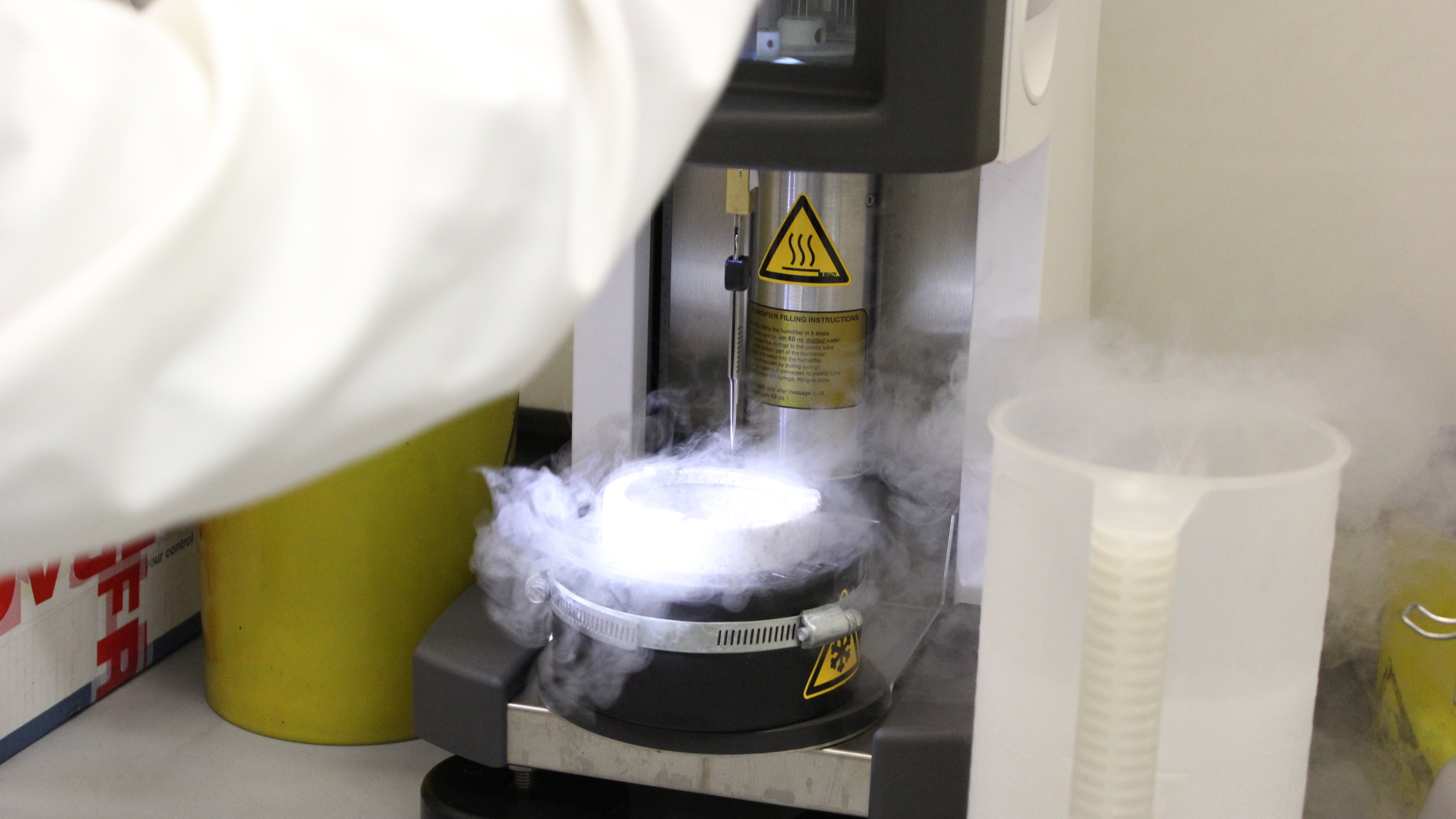
Image: The ICR's cryogenic electron microscopy (cryo-EM) facilities in Chelsea, London.
Scientists have successfully used a new imaging technique to determine the structure and interactions of a protein complex that plays a significant part in the initiation and progression of cancer. They showed that it was possible to use a high-resolution form of cryogenic electron microscopy (cryo-EM) to gain a better insight into how the protein complex binds to inhibitor drug molecules.
The protein complex – cyclin-dependent kinase (CDK)-activating kinase, known as CAK – has shown promise as a target for cancer drugs, so a full understanding of its molecular composition should facilitate the structure-based design of new, effective treatments.
In addition, other researchers will be able to apply this new methodology to their own work, so it has potential to aid drug discovery very broadly, both geographically and across cancer types.
The study was led by researchers at The Institute of Cancer Research, London, who collaborated with groups at Imperial College London and Thermo Fisher Scientific. It was largely funded by The Institute of Cancer Research (ICR), which is both a research institute and a charity, with additional funding coming from Cancer Research UK, Thermo Fisher Scientific and the Medical Research Council of the UK. The paper has been published in the journal Nature Communications.
Applying cryo-EM to structure-based drug discovery
Cryo-EM is a cutting-edge technique that reveals the detailed structure of biological molecules by rapidly freezing samples to below -150°C and then passing a beam of electrons through them to collect images from different angles. These images go to a computer, which uses them to create a three-dimensional model of the molecules.
Although cryo-EM is a useful tool in structural biology, the resolution of the images it produces can be limiting, and it cannot always process sufficient information for researchers to get the detail they need within a reasonable time frame.
Faced with this challenge, the ICR-led team decided to employ an improved cryo-EM workflow. The researchers began by performing rapid screening using a less costly and more readily available cryo-EM instrument, which requires the specimen to be in a vacuum. They assessed the resulting images while the data collection was still ongoing and progressed only the specimens with high potential to the next stage. Here, they used a piece of technology called a cold field emission gun, which provides higher resolution images and is ideally suited for obtaining detailed views of small molecular complexes.
This approach enabled the researchers to achieve exceptional images, with the majority at 2 angstrom (Å) resolution. Prior to this work, such a high resolution had only ever been achieved for one protein complex of a similarly small size, and it was not a medically relevant target. The team also showed that it was possible to determine more than 12 structures of up to 4 Å resolution a day by using a high-specification microscope.
Building knowledge about CAK
CAK is a protein complex made up of CDK7, cyclin H and MAT1. It is responsible for activating several other CDKs, which regulate the cell cycle and ensure the accurate replication of DNA during cell division. If these CDKs are activated abnormally, this can lead to the uncontrolled growth of cancer cells. Due to this, CAK has been identified as a promising therapeutic target in cancer.
Six experimental drugs that target CAK by inhibiting CDK7 have been progressed to clinical trials, but the development of these compounds is very challenging because the structure of the active site on different CDKs is very similar. This makes it difficult to target CDK7 without also targeting other CDKs.
In this study, the researchers determined 16 detailed structures of the catalytic module of CAK, which is the part that facilitates its reactions. These included structures both of CAK in its free state and of CAK when bound to other substrates. Based on these structures, the team was able to reveal certain mechanisms of CDK7 selectivity that should lay the foundations for the future design of CDK7 inhibitors.
“Broadening the scope of its impact”
First author Victoria Cushing, a PhD student at the ICR, said:
“This methodological advance is really exciting for the structural biology community. It has overcome a longstanding technical challenge, allowing us to achieve both high resolution and high throughput. We have already used it to provide detailed insight into the interactions between CAK and its inhibitors, which should support the design of next-generation therapeutics.”
Senior author Dr Basil Greber, Group Leader of the Structural Biology of DNA Repair Complexes group at the ICR, said:
“We are excited to have established a framework for the application of cryo-EM to structure-based drug design. It will be particularly useful for small target proteins that cannot be crystallised, for which structure-based methods were previously inaccessible. We hope that our work will enable scientists in other labs and institutions or in industry to apply this methodology to drug discovery, broadening the scope of its impact considerably.”