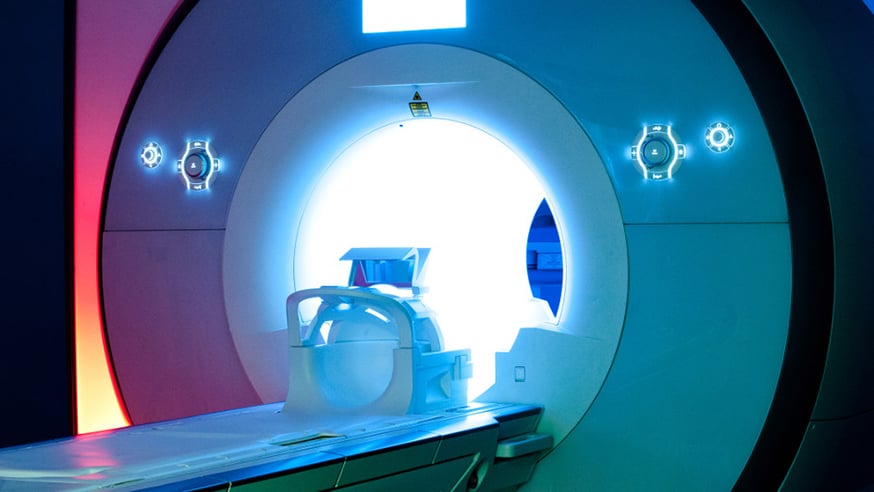
An MRI scanner
A magnetic resonance imaging (MRI) technique that can measure tumours in the body could be an easy and reliable way to assess lung cancer’s response to treatment, a new study shows.
The study paves the way for clinical trials of the technique in lung cancer – and could ultimately allow doctors to take more accurate scans than are currently possible.
Scientists at The Institute of Cancer Research, London, and The Royal Marsden NHS Foundation Trust led the study, which involved taking scans, twice over, from 23 patients treated at four different hospitals.
The researchers measured the effect of using different types of software to process the data produced during the scans – an important consideration because different hospitals use different technology.
The study was published in the journal European Radiology, and was supported by the EU Innovative Medicines Initiative, with funding from Cancer Research UK and the Engineering and Physical Sciences Research Council to the Cancer Imaging Centre at the ICR and the Royal Marsden.
The ICR's Magnetic Resonance team develops and tests new probes, instrumentation and techniques to better plan and assess cancer treatment.
Consistent results
In the study, the researchers measured the movement of water in and out of tumour cells during MRI scans – using a measurement called the Apparent Diffusion Coefficient.
The study found that measurements varied by less than 10% between scans across all patients’ tested and were consistent across hospitals – not affected by where the scans were taken or the equipment and software used.
For tumours larger than 3cm in diameter there was less than 4% difference between ADC values. Measurements of smaller tumours, which are more affected by movement from breathing, still showed a high degree of repeatability, with a maximum variation of less than 10% between scans.
Importantly, patients could breathe freely during scans – which is often not possible during scans taken for routine care at the moment.
Study leader Professor Nandita deSouza, Professor of Translational Imaging at the ICR and Honorary Consultant at The Royal Marsden, said: “Measuring the apparent diffusion coefficient of lung cancer with MRI is both convenient and non-invasive, but this is the first time research has shown ADC values can be reliably measured using repeat scans in a multi-centre setting.
“Our study shows measuring changes in ADC for lung cancer is manageable for patients, with comparable results using different MRI. Importantly it proves that we can proceed with multi-centre lung cancer trials despite differences in the technology between hospitals.”
