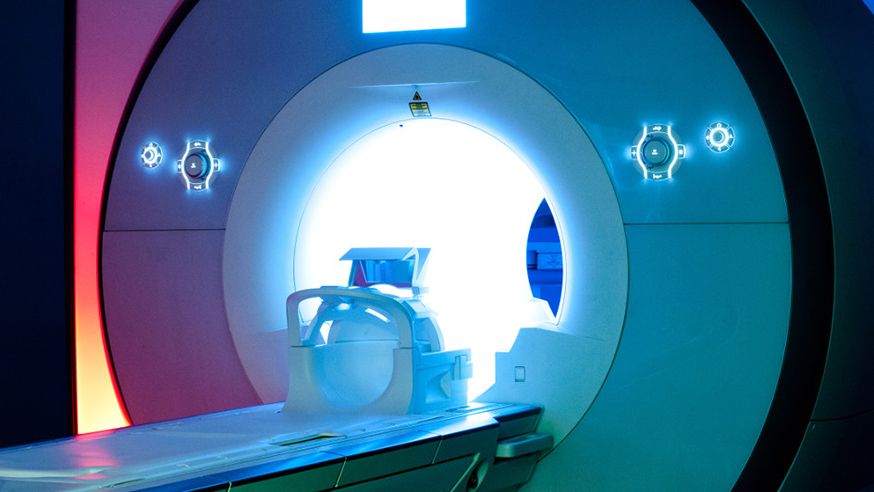
An MRI scanner at The Royal Marsden NHS Foundation Trust
A combination of functional imaging techniques can help determine which regions of a brain tumour are likely to spread undetected, according to a new study.
High-grade brain tumours can vary greatly in terms of their growth, density, quality of new blood vessels, and how they spread. But using imaging to accurately inform on these processes remains difficult.
In this study — published in NMR in Biomedicine — a team of scientists from The Institute of Cancer Research, London, and The Royal Marsden NHS Foundation Trust investigated using a combination of magnetic resonance imaging (MRI) techniques to get a clearer picture of brain tumour growth.
The researchers used MRI scans with two different types of small magnetic particles — ultrasmall superparamagnetic iron oxide (USPIO) and gadolinium containing Gd-DPTA — that act as contrast agents to help sharpen the difference between tissue and blood vessel.
A clearer picture
If Gd-DPTA begins to leak into the tissue from the blood, particularly in the brain it indicates that the blood vessels are immature and weaker than normal healthy blood vessels that maintain the blood brain barrier.
This is typical of new blood vessels created when tumours require more nutrients.
By contrast, USPIO gives a better indication of the overall network of blood vessels, highlighting where tumours are co-opting local blood vessels rather than signalling for the creation of new ones.
In Europe, only gadolinium-based contrast agents are in use in the clinic, but as this only highlights areas of new blood vessel formation it may not show the entire tumour volume.
By analysing these data in conjunction with other MRI biomarkers, such as the ability of water to spread through the tissue, the researchers were able to create a far deeper insight into the overall state of the vasculature around the tumour.
The researchers tested this imaging strategy on mice with two distinct brain tumours and successfully showed the different growth patterns and blood vessel formation of each.
First study of its kind
While these techniques have been tried in parallel in other preclinical studies, this is the first time that they have all been analysed together spatially — displaying which areas of the tumour are likely to be growing and gathering new blood vessels.
Understanding which regions of a brain tumour are likely to spread is essential for clinicians planning treatment, as this can guide radiotherapy and surgery.
Lead author Dr Jessica Boult, located within the ICR’s Magnetic Resonance Team, said: “High-grade brain tumours desperately need new treatments. And one of the biggest challenges facing doctors is being able to tell the difference between tumour and healthy tissue where the blood brain barrier is intact.
“In the future, such techniques can help clinicians more accurately plan surgery and radiotherapy as well as monitor tumour therapy response.”
