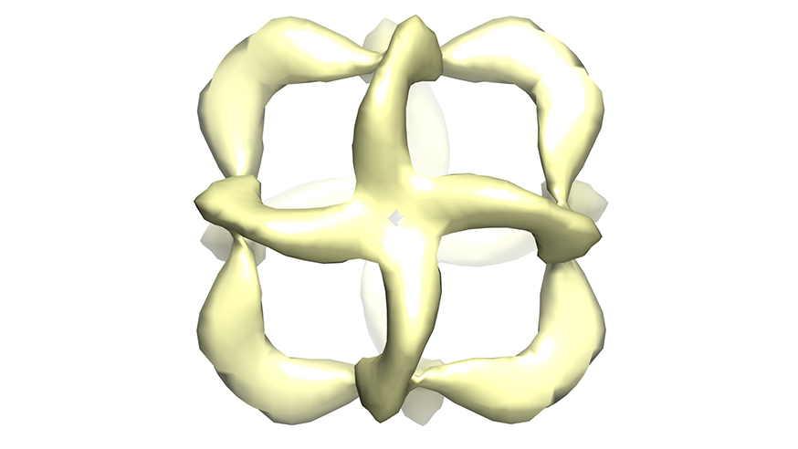
A 3D model from electron microscopy data of an octahedral nanostructure composed entirely of artificial XNA polymers (image: A.Taylor, F. Beuron, S-Y. Peak-Chew, E.P. Morris, P. Herdewijn, P. Holliger)
Scientists have built the first 3D nano-sized objects using artificial DNA, which could be used to deploy cancer treatments inside tumour cells.
The teams from The Institute of Cancer Research, London, and the Medical Research Council (MRC) Laboratory of Molecular Biology created microscopic pyramid- and diamond-shaped 3D ‘packets’ by folding together artificial nucleic acid building blocks called Xeno nucleic acids (XNAs).
They saw that XNA made pyramid packets that were more stable in biological environments than DNA-made structures, keeping their shape for eight days compared with DNA nanostructures, which degraded after two.
The research was published in ChemBioChem and funded by the Medical Research Council and the Biotechnology and Biological Sciences Research Council, with additional support from Cancer Research UK and the European Science Foundation.
DNA nanotechnology is an exciting new way to manipulate genetic material, which could have huge benefits for biomedical research and clinical care.
Strands of DNA or RNA can be folded to make microscopic packets which could detect important biological markers of cancer, or be used to transport cancer treatments into cells to make them more effective. However, DNA-cutting enzymes in human tissue can break down these nanostructures very rapidly, limiting their use as medical treatments.
The 3D nanostructures using XNA building blocks – in which the sugar backbone of human nucleic acid strands is chemically altered in ways that do not occur in nature – were first developed by the University of Cambridge and had greater bio-stability and wider ranging physical and chemical properties than DNA.
'Great promise'
They saw that XNAs behaved in a similar way to DNA nanostructures, folding to produce 3D tetrahedrons (pyramid shaped) and octahedrons (diamond shaped) as intended, which ICR researchers confirmed using electron microscopy.
When tested inside cell cultures, tetrahedral XNA packets kept their shape inside for eight days, compared with structures made from DNA which degraded after two days.
Dr Edward Morris, Leader of the ICR’s Structural Electron Microscopy team, said: “DNA has shown great promise as a potential building material for nano-molecular scale objects, but unfortunately they tend to get broken down quite quickly by our bodies and this may limit their clinical use. Our research with scientists from the MRC Laboratory of Molecular Biology shows that you can make robust microscopic 3D shapes using this novel XNA chemistry which can stand up to conditions inside the body.
“Using our expertise in electron microscopy, we saw that synthetic XNA folded to make 3D-shaped nanostructures which correspond closely to analogous structures obtained with conventional nucleic acids such as DNA. The findings are an important first step for this exciting new genetic construction technique, which could lead to new ways to target cancer treatments directly at cancer cells.”