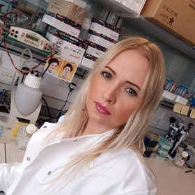
Preclinical Molecular Imaging Group

Dr Gabriela Kramer-Marek’s group uses cutting-edge biomedical imaging techniques to gain information about the way particular genes drive cancer progression.

Our group’s long-term goal is to develop specific biomarkers for detecting cancers and to evaluate these biomarkers in pre-clinical cancer models

Notwithstanding the remarkable clinical success of mAb-based treatment regimens, not all patients benefit from them. This can be attributed, at least in part, to the complexity of the tumour microenvironment and its considerable heterogeneity both in terms of the tumour and non-tumour cell components. These phenomena represent a huge challenge in identifying predictive biomarkers and stratifying patient populations for personalised therapy approaches.

Therefore, there is an urgent need to develop assays that will help in three ways:

  1. accurate patient selection
  2. understanding intrinsic resistance mechanisms or the emergence of acquired resistance following treatment initiation and
  3. choosing the most effective combination regimen in circumstances in which single-agent therapies are insufficiently effective.

Currently, the baseline expression level of antigens targeted by therapeutic mAbs can be analysed by methods such as: immunohistochemistry (IHC), flow cytometry, proteomics, or next-generation sequencing of tumour tissues acquired at diagnostic biopsy or intra-operatively. These techniques aid our understanding of how cancer cells adapt to treatment and become resistant, but such methods are inherently invasive, prone to sampling errors caused by inter- and intra-tumour heterogeneity of receptor expression within analysed biopsy specimens and do not lend themselves readily to repeated sampling.

Positron emission tomography (PET), using radiolabelled mAbs, antibody fragments or engineered protein scaffolds (immuno-PET), has the potential to acquire information non-invasively and can be highly complementary to analyses based on tissue acquisition. Accordingly, immuno-PET agents might accurately identify the presence and accessibility of the target and provide a rapid assessment of tumour response to a variety of treatments in a timely fashion (e.g. within 1-2 weeks of treatment initiation).

Furthermore, immuno-PET agents can provide information about the heterogeneity of both target expression and therapeutic response, which are increasingly recognised as key factors in treatment resistance. This especially relates to patients with advanced disease in whom target expression may vary from site to site and a biopsy of a single local or metastatic deposit may not accurately reflect the situation across the entire disease burden. Although introduction of immuno-PET into routine clinical practice may add complexity and increase costs, with appropriate use this imaging modality has the potential to identify patients likely to benefit from therapy and assess the efficacy of novel target-specific drugs.

Against this background, our research focuses on the development and characterisation of targeted-PET radiotracers, including protein-based theranostic agents that enable smart monitoring of immunotherapies and expand opportunities for personalised medicine approaches.

Early diagnosis and individualized therapy have been recognized as crucial for the improvement of cancer treatment outcome. While proper molecular characterization of individual tumour types facilitates choice of the right therapeutic strategies, early assessment of tumour response to therapy could allow the physicians to discontinue ineffective treatment and offer the patient a more promising alternative. Therefore, the role of molecular imaging in elucidating molecular pathways involved in cancer progression and the ability to select the most effective therapy based on the unique biologic characteristics of the patient and the molecular properties of a tumour are undoubtedly of paramount importance.

The mission of this group is to investigate innovative imaging probes and apply them to novel orthotopic or metastatic models that are target driven, to gain information of the way particular oncogenes drive cancer progression through signalling pathways that can be imaged in vivo and, correlate it with target level ex vivo. Such an approach enables non-invasive assessment of biochemical target levels, target modulation and provides opportunities to optimize the drug dosing for maximum therapeutic effect, which leads to the development of better strategies for the more precise delivery of medicine.

The long term goal of our research is to develop specific imaging cancer biomarkers, especially for positron emission tomography (PET) as well as optical imaging and, evaluate these biomarkers in pre-clinical cancer models. Significant efforts are directed towards validating biomarkers for early prediction of treatment response, with the focus on new targeted therapies (such as inhibition of cell signalling pathways).

Our initial portfolio of imaging agents include radiolabelled affibody molecules, TK inhibitors and, conventional tracers that monitor universal markers of tumour physiology.

We are actively supported by other groups from the Division of Radiotherapy and Imaging as well as the Division of Cancer Therapeutics. Moreover, our close association with The Royal Marsden NHS Foundation Trust enables rapid translation of our research to early clinical studies and ensures a fast transition of know-how from the research laboratory to the patient bedside.

Dr Gabriela Kramer-Marek

Group Leader:

Preclinical Molecular Imaging Gabriela Kramer-Marek

Dr Gabriela Kramer-Marek is investigating new ways of molecular imaging in order to predict an individual patient’s response to treatment. Before moving to the ICR, she developed a new approach for non-invasive assessment of HER2 expression in breast cancer.

Researchers in this group

.

Phone: 020 3437 6376

Email: [email protected]

Location: Sutton

.

Phone: +44 20 3437 6785

Email: [email protected]

Location: Sutton

.

Phone: +44 20 3437 6857

Email: [email protected]

Location: Sutton

.

Phone: 020 3437 4549

Email: [email protected]

Location: Sutton

Dr Gabriela Kramer-Marek's group have written 63 publications

Most recent new publication 10/2024

See all their publications

Recent discoveries from this group

17/02/25

Scientists have shown for the first time that some of the most persistent cancer cells in glioblastoma, a common type of adult brain tumour, rely on a specific enzyme for survival and that inhibiting this enzyme leads to the death of the cells.

Furthermore, they have demonstrated that it is possible to reprogram other cancer cells to become dependent on this enzyme – called transforming growth factor beta-activated kinase 1 (TAK1) – which would mean that TAK1 inhibition could destroy more of the tumour and potentially prevent a recurrence of the disease.

This early-stage research provides hope that TAK1 could be an important therapeutic target, not only in glioblastoma but also in other types of cancer.

The study was led by researchers at The Institute of Cancer Research, London, and published in the Nature journal Cell Death & Disease. The work was primarily funded by the Neye Foundation and the Brain Tumour Charity, with additional financial support provided by the Danish Cancer Society, Memorial Sloan Kettering Cancer Center and Cancer Research UK. The Institute of Cancer Research (ICR), which is both an institute and a charity, also contributed to the study’s funding.

Specific challenges in treatment development

Even though glioblastoma is the most common form of primary brain tumour in adults, the treatment options remain limited because the disease is so difficult to treat.

This is partly because the tumours are often detected at a late stage, but there are also various biological factors at play. Firstly, there are typically significant differences – relating to shape, genetics and behaviour – between glioblastoma cells, both across patient populations and within the same tumour. Secondly, the position of the cancer in the brain means that treatments can only reach it if they can penetrate the blood-brain barrier.

These difficulties are compounded by the poor response of glioblastomas to most cancer medications, which tends to result in tumour recurrence. Previous work has attributed this treatment resistance to a persistent subset of cells called glioblastoma stem cells (GSCs), which can be divided into three subtypes. The mesenchymal subtype correlates with the poorest patient outcomes.

Earlier studies have also demonstrated that glioblastoma cells can shift between subtypes and that a transition to a primarily mesenchymal cell state occurs in many of the patients whose disease relapses following treatment. In this new study, the researchers therefore aimed to identify a potential vulnerability in mesenchymal glioblastoma cells that could be exploited therapeutically.

Revealing a selective dependency

The researchers used a method called Clustered Regularly Interspaced Short Palindromic Repeats (CRISPR) screening to look for genes that help cancer cells survive. They adopted an unbiased approach, investigating the effects of inactivating the function of thousands of genes rather than limiting themselves to genes already known to have a role in cancer cell survival.

They discovered that the survival of many GSCs is dependent on the activity of TAK1, which seems to inhibit receptor-interacting protein kinase 1 (RIPK1). When active, RIPK1 associates with a protein complex called the caspase-8/FADD complex to induce cell death. By blocking RIPK1’s activity, TAK1 prolongs the survival of the cancerous cells.

The team was also able to identify a genetic ‘signature’ that could support treatment decisions by revealing which tumours are likely to respond well to drugs that inhibit TAK1. This signature was present in cells with high levels of immune activation, including those with a mesenchymal subtype, and the team successfully used it to predict sensitivity to a known TAK1 inhibitor.

The researchers then took their work a step further, showing that it was possible to reprogram other cancer cells to become dependent on TAK1 function for their survival. They did this by treating the cells with specific cytokines – substances made by immune cells – to mimic an immune-cell rich environment. This reprogramming serves to make a larger number of cells sensitive to treatment with TAK1 inhibitors.

“Half of all glioma patients might benefit”

First author Dr Helene Damhofer, who was a staff scientist at the ICR at the time of the study and is now a senior scientist at BiOrigin in Copenhagen, said:

“This innovative work has provided a link that was not known before. By discovering that a subset of glioma cells with a high level of immune activation is dependent on TAK1, we have provided insight into the mechanism by which TAK1 inhibition induces cell death in the glioma cells. Excitingly, this also gives us a means by which to stratify patients, as we can predict those who will likely benefit from TAK1 inhibition.”

Senior author Professor Kristian Helin, CEO of the ICR and Group Leader of the Epigenetics and Cancer Group in the ICR’s Division of Cancer Biology, said: 

“We are pleased to be the first to show that several cancer types are inherently dependent on the function of the kinase TAK1 to suppress cell death. The findings of our study suggest that half of all glioma patients might benefit from TAK1-targeting therapy.

“What’s even better is that the mechanisms of TAK1 dependency that we’ve uncovered are not limited to glioma cells. We had not anticipated that these mechanisms would be preserved against so many cancer types, but it means that many more people with cancer might benefit from treatments that inhibit TAK1 in the future.”

The team is optimistic that this study is just the first step towards effective new cancer treatments. Dr Damhofer said:

“We hope our work inspires other cancer researchers to test TAK1-targeted therapies in their model systems and find optimal combination therapies. Moreover, we hope it will inspire biotech and pharma companies to develop TAK1 inhibitors that can penetrate the blood-brain barrier. Ultimately, we believe that a TAK1 inhibitor could enhance treatment options for patients with glioblastoma or other tumours showing an immune-activated gene expression signature.”