Flying bats, war-time submarines, pictures of unborn babies and cutting-edge cancer treatment – what do these things have in common? The answer is ultrasound, an incredibly versatile form of energy that has been applied to navigation, locating objects, medical imaging and diagnosis, and is now at the forefront of treating cancer.
In 1794, it was the physiologist Lazzaro Spallanzani who was the first to study echolocation among bats, which formed the basis for ultrasound physics. But it wasn’t until 1942, that neurologist Karl Dussik was the first to use sonography for medical purposes, when he transmitted an ultrasound beam through the human skull in an attempt to detect brain tumors.
Ultrasound has since gone on to find a wide range of applications in engineering and medicine. The two major uses of ultrasound in the clinic are for imaging and treatment. At first glance these applications seem on opposite poles. Medical imaging – think about those prenatal scans for which ultrasound is best known – requires the gathering of information from tissue, while successful therapy requires heating up and destroying target tissues. But it is the versatility of ultrasound, and the fact that it can be so tightly controlled, which makes it such an attractive option for cancer therapy.
Professor Gail ter Haar, Professor of Therapeutic Ultrasound here at The Institute of Cancer Research in London has been one of the pioneers in developing the ultrasound technology used in high-intensity focused ultrasound (HIFU) – where ultrasound energy is focused and used to selectively target tumour tissue. She recently rounded up the current, and possible future applications of using ultrasound as therapy in a review published in the International Journal of Hyperthermia.
I spoke to Professor ter Haar about where she sees therapeutic ultrasound going next, and what area she is particularly interested in. She told me: “Ultrasound is an exciting, versatile technique. Many researchers are now undertaking pre-clinical research, so we can expect lots of new treatment techniques to emerge. Using HIFU to treat cancer is rapidly gaining widespread clinical acceptance, and we are extremely proud to be at the forefront of its development.”
Upon reading her review, I found it fascinating how ultrasound can be so versatile – it can be used to image unborn babies, but can also obliterate tumours. Professor ter Haar explains in her review that it is the differences in the intensity of sound waves which can deliver ultrasound’s varied applications. The Ultrasound energy used in pregnancy scans ranges from 0.01–0.1 W/cm2, whereas in HIFU the range is from 800–1500 W/cm2 – nearly 10,000 times more powerful.
It’s this continuous high intensity of waves used in HIFU that creates tissue heating, arising from friction as tissues absorb sound energy. When short pulses at low repetition rates are used, it is possible to avoid thermal effects and call into play ‘mechanical’ effects instead. One of the most disruptive mechanical effects is a phenomenon called acoustic cavitation, where small bubbles of gas (or microbubbles) form.
In the past, clinicians have normally tried to avoid the formation of microbubbles when using ultrasound as they can damage tissue in an unpredictable way. But many of the therapeutic techniques currently under investigation purposely introduce microbubbles. An area that I found particular interesting was the fact that microbubbles can be used to open up the blood-brain barrier, a tightly packed layer of cells that line the brain’s blood vessels. This impenetrable barrier shields the brain from infections, toxins, and other threats, but also prevents therapeutic drugs from being able to access the brain. If researchers can temporarily open up the barrier safely, and in a controlled manner, it could overcome a significant hurdle in treating brain cancer.
Taking microbubble activity to the extreme, scientists have developed a technique called histotripsy. Here, the ultrasound pressures used are hundreds of times higher than regular diagnostic imaging, at a similar level to that used to break down kidney stones. Although this sounds frightening, it can be tightly controlled and can remove tissue with a very sharp boundary. Histotripsy has already been used in the clinic to remove cardiac tissue in treatment of congenital heart disease, and could be used in the treatment of prostate cancer. But there is still much work to be done in optimising treatment delivery and monitoring.
Researchers are also exploring the potential of using the thermal effects of ultrasound for increasing drug delivery. Drugs can be incorporated into vehicles with temperature-sensitive shells that only release their contents when they reach a part of the body that’s at a temperature that disrupts them. The ability of ultrasound to heat target tissues within the body very selectively makes it perfect for achieving this local drug release.
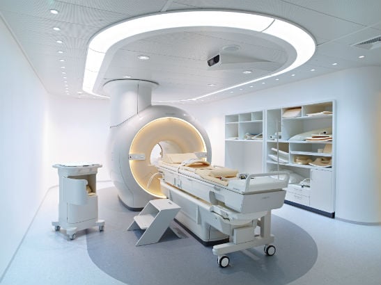
As for future research directions for ultrasound at the ICR, Professor ter Haar is now collaborating with Professor Nandita deSouza on a clinical trial that uses magnetic resonance image-guided HIFU to alleviate cancer pain by destroying the nerve tissue in the bone around the tumour.
“Early results in patients with bone cancer have so far been promising,” says Professor ter Haar. “Using this non-invasive technique in patients for whom radiotherapy is no longer an option – or where other treatment avenues have been unsuccessful – will offer hope where all else has failed.”
Ultrasound as a medical application has been with us for more than 75 years, but it seems that it is only now that are we exploring its full potential for improving cancer treatment.
To read more about HIFU use in bone cancer, you can watch our video and press release by clicking here.
For more background, you can read our feature on HIFU by clicking here.
