Computational Pathology and Integrative Genomics Group
The group works to reconstruct two-dimensional or three-dimensional tumour models from images of tumour sections. Previously led by Professor Yinyin Yuan, it is now led by Professor Janet Shipley as interim.
Our group is investigating cancer tissue ecosystems and how selective pressures can promote cancer development.
Deciphering Tumour Microenvironement
A pathologic-genomic approach
Pathological analysis of tumour architecture and cell morphology dates back to the 19th century. Now, morden image-analysis tools promises objective and quantitative insights into hundreds of tumours.
By drawing on the rich information from pathological images and combining it with molecular data from next generation sequencing, we hope to offer more powerful approaches to study the microenvironment and its implications to cancer progression.
Building an automated classifier for histopathological images, enables us to identify heterogeneous cell populations in patient tumours and quantify their spatial proximity to each other.
Quantitative scores of cell spatial patterns
The cell spatial patterns can be summarised using methods from spatial statistics and pattern recognition techniques to provide quantitative scores.
These quantitative scores of cell patterns can reveal various metrics that would be impossible to conclude by eye, for example, the degree of clustering and the spatial interactions between different cells.
Discovery of functional roles of normal cells
One of the central dogma in cancer biology is the functional roles of heterogenous types of cells in cancer progression.
With quantitative readout from the images, we can correlate the abundance and spatial patterns of cells with patient outcome. For example, by summarising the abundance of lymphocytes in the ER-negative breast cancer, we are able to stratify patients into different outcome groups, where the patients with high numbers of lymphoctyes have significantly better prognosis than patients with few lymphocytes.
Integrating images with molecular signal
Combining molecular signal from the same samples, the image-based quantitative scores can either complement molecular data, or offer completely new observations from this entirely different angle.
The Computational Pathology and Integrative Genomics Team sits within the Centre for Evolution and Cancer and the Centre for Molecular Pathology. The scientists within this team are investigating cancer tissue ecosystems and how selective pressures can promote cancer development. Understanding how tumours adapt to their microenvironment is key to appreciating how they grow and spread, and could direct interventions to alter or stop the selective pressures helping to drive their growth.
As cancer cells all evolve dynamically within the habitats of our tissues on which they retain some dependence, therapeutic regulation of the ecosystem may be a viable alternative to the current notion of trying to kill cancer cells directly - a bit like draining the swamps to get rid of mosquitoes as one way of eradicating malaria.
Such approaches might provide a Darwinian bypass that uses selective pressure on the tumour ecosystem to avoid the problems that drug resistance can bring. In fact doctors can already target the ecosystem, with anti-hormonal therapies (such as tamoxifen for breast cancer), anti-inflammatories (such as aspirin for gastrointestinal cancer), and anti-angiogenesis therapies (that stop new blood vessel formation) are already being used for this.
The team focusses on developing computational approaches to combine statistical modelling with automated image analysis of tumours. The scientists are looking at the different regions within a genetically variable (heterogeneous) tumour and analysing them as if they are different ecological systems.
The team trains powerful computers to automatically identify cancer cells in pathology samples from the clinic. By drawing on the rich information contained in these images, and combining it with the data describing the DNA sequences of the cancer cells, so the team can construct two and three dimensional images of tumour sections. This enables them to explore the complexities of the tumour microenvironment and to develop new tools to diagnose patients or to predict how they might respond to a treatment, and to explore the consequences of how different regions and populations of cells within the tumour interact.
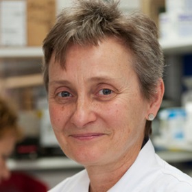
Professor Janet Shipley
Group Leader:
Sarcoma Molecular Pathology, Computational Pathology and Integrative Genomics
Professor Janet Shipley is investigating ways to improve the treatment of patients with sarcomas that have a poor outcome.
