Clinical Freehand Reflection-Mode Photoacoustic Imaging
M Jaeger, DD Birtill, A Gertsch, JC Bamber, in collaboration with E O’Flynn, Radiology Department and CRUK-EPSRC Cancer Imaging Centre.
Source of funding: CRUK, EPSRC, Institute of Cancer Research, EU Marie-Curie, Zonare Medical Systems Inc
Photoacoustic (PA) imaging is an emerging imaging modality offering promise for cancer diagnosis, therapy planning, and monitoring response. The images show optically absorbing tissue structures based on detection of ultrasound signals after pulsed laser illumination.
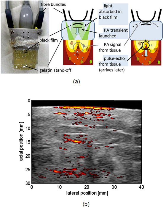
For clinical use, PA imaging is best implemented with freehand pulse-echo scanning in a single device, so that PA signals may be displayed in real-time within the anatomical context. For this purpose, PA frames must be interleaved with pulse-echo frames. To overcome the fact that this is normally not feasible with commercially available ultrasound imaging systems, and for clinical research purposes, we have developed a novel approach where both PA and pulse-echo images are generated photoacoustically. This was implemented on a commercial scanner (Z.one™ Zonare, Mountain View), using a Q-switched Nd:YAG laser at 1064 nm wavelength and 10 Hz pulse rate. Each laser pulse results in a data frame from which both a PA and a PA-echo image is reconstructed; PA signals are generated directly from optically absorbing tissue structures, whereas a transmit pulse for echography is generated by a black absorbing film placed inside an acoustic stand-off that also permits temporal separation of the two types of information.
Preliminary clinical evaluation suggests that although echograms generated with a PA transmit pulse were of poorer resolution and were noisier than those obtained using a focused transmit pulse, they correspond well to conventional pulse-echo images, making them a valuable surrogate and permitting registration of PA images with high quality echograms (Figure 9). Strong PA signals were obtained from the skin and tissue structures to depths > 2 cm. PA clutter appears to limit clinical image quality, particularly at large depths, and work is underway to develop successful clutter-reduction strategies.
 Fig.9. (a) Photograph of the scan head with the photoacoustic (PA) stand-off, and diagrammatic illustration of principle of operation for combined PA and PA-echo imaging. (b) Example preliminary Duplex thresholded PA (colour) and conventional B-mode (grey scale) image of a breast tumour. A PA-echo image (not shown) was used to register the PA data with a conventional high quality B-scan, obtained later.