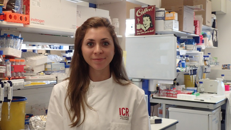
ICR PhD student Laura Mariotti
Doing a PhD in academic research is like solving a giant puzzle: placing one piece at a time, making sure it fits and seeing a pattern emerge.
I’m a final-year PhD student in the Division of Structural Biology at The Institute of Cancer Research, London. We’re interested in finding out what a protein looks like – its three-dimensional shape.
Once we know what a protein looks like, it helps us to understand a protein’s function and how, in cancer cells, these proteins can go wrong. We can determine which parts or specific atoms of the proteins are important for their roles in normal and cancer cells. This is valuable information for chemists, who might design drugs that turn the protein on or off.
But finding out a protein’s shape is challenging, in part because of the small size of the molecules we’re interested in: proteins have a diameter of a few nanometres (1 nanometre is 10-9 m). To give you an idea of how small they are, a sheet of paper is approximately 100,000 nanometres thick.
Seeing individual atoms
We use two main techniques in structural biology, called protein X-ray crystallography and electron microscopy.
For protein X-ray crystallography, we make a crystal of our protein of interest – many millions of copies of the protein, all slotting together in a highly ordered way.
Then we irradiate the crystal with X-rays to create a map, which we call a diffraction pattern. From this map, we can deduce the position of the atoms within the protein.
In electron microscopy, we image our protein at very high magnification (around 50,000x) using a beam of electrons, rather than light. We make two-dimensional projections of our protein in many different orientations, and use these different views to re-construct a model of the 3D object.
Studying a target
My team focuses on a protein called Tankyrase, which controls many processes that are vital for the growth and function of cancer cells.
Tankyrase is an enzyme, performing a chemical reaction in a cell where it modifies proteins – usually targeting other proteins for degradation. It is part of several signalling cascades, which are sequences of events that lead to a specific cellular outcome, such as a cell growing or dividing.
Some of these signalling cascades are distorted in cancer, leading to the wrong outcome in a cell – uncontrolled growth, for example. However, the cancer cells become dependent on these signalling cascades to survive, and this vulnerability can be exploited by targeting proteins like Tankyrase.
Tankyrase is composed of different elements. One part of Tankyrase recognises and binds other proteins, which are known as substrate proteins. Another part adds a molecular tag onto these substrate proteins. This tag is usually a signal for other proteins to target the substrate proteins for degradation.
A third entity binds Tankyrase molecules together to create a ‘string’ which acts as a platform in the cell, recruiting and modifying many substrate proteins at once.
The main aim of my PhD project is to determine the 3D shape of the ‘string’ formed by multiple Tankyrase molecules and understand its role.
It’s a team effort – an aspect of academic research that I particularly like.
Lab vs office
I enjoy working both at a desk and at a lab bench. Every week, I need to find the right balance between carrying out experiments in the lab and analysing the results in the office.
In my team, we have weekly lab meetings where we each, in turn, explain the experiments we did that week and the results obtained. These conversations are incredibly useful.
I have one-to-one meetings with my supervisor, which is a great way to make sure I’m still heading in the right direction. Every couple of months, I also present my work in front of a bigger audience in a slightly more formal setting. These presentations are a great opportunity to put my results into figures, receive feedback and think about my project from a broader perspective.
Equipped for the future
I will start writing my PhD thesis soon. Summarising three-and-a-half years’ worth of work in a manuscript will be an exciting challenge. I also need to think about my next adventure, which could be in academia, industry or even something else.
No matter what I choose to do next, studying for my PhD at the ICR has equipped me with all the skills I need for the future and has opened many doors.
comments powered by