Image guided Theranostics in Multiple Myeloma (iTIMM)
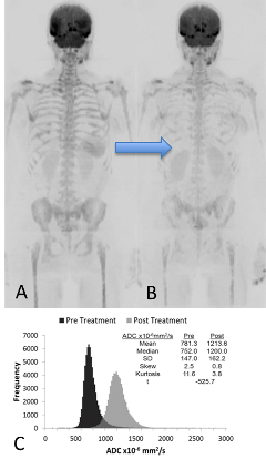
We have demonstrated that whole body diffusion weighted magnetic resonance imaging (MRI) is an extremely sensitive tool for detecting disease within the bone marrow. In addition our previous studies have shown the capability for whole skeleton quantitative measurements of disease burden and response to treatment (Figure 1, right).
The iTIMM study is investigating the potential of whole body diffusion weighted MRI for detection of minimal residual disease following autograft with a view to using this imaging to guide adaptive treatment regimens.
We will also explore the relationships between quantitative measures of disease phenotype, burden and response with outcomes and standard measures of risk such as cytogenetics.
Figure 1 (right) - Whole body diffusion weighted magnetic resonance imaging of a skeleton
Pre-operative Imaging in Retroperitoneal Sarcomas (PIRS)
This is a prospective multiparametric MRI study of tumour heterogeneity. We are establishing reproducibility of functional MRI parameters and defining their relationships to histology.
Understanding which parameters are robust and reflect aggressiveness allows three-dimensional maps of tumour heterogeneity to be built using supervised machine learning techniques for local treatment planning and response assessment. For patients who have undergone radiotherapy we are investigating the interaction between functional MRI parameters to produce quantitative and visual tools which reflect heterogeneous response. This study will open up new opportunities for image guided adaptive therapy and has been performed in close collaboration with the soft tissue sarcoma unit at The Royal Marsden NHS Foundation Trust.