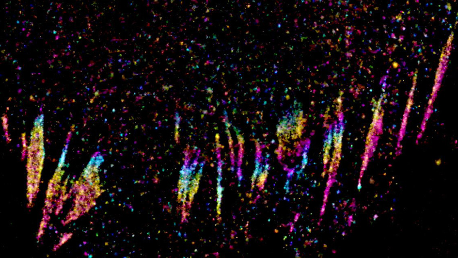
Image: Super-resolution microscopy image of cell focal adhesions by PhD Student Ian Jones, Dr Lucas Dent, and Professor Chris Bakal
Metastasis, when cancer moves and spreads around the body leading to the development of secondary cancer growths, is the cause of about 90% of all cancer deaths.
It happens because cancer develops the pernicious ability to escape the tissue where it formed and invade healthy tissue elsewhere. And once cancer has spread it’s almost always fatal.
Cells assume a wide variety of complex shapes in order to carry out different roles in the body, and metastatic cancer cells are able to manipulate their own shape to help them spread.
Professor Chris Bakal, Professor of Cancer Morphodynamics at The Institute of Cancer Research, London, leads a team examining the genetic and biochemical mechanisms that underpin these shape changes, and their role in cancer.
His team use state-of-the-art imaging equipment to study the biological switches that cause cells to change shape, become cancerous and spread around the body.
Catching cancer in the act to understand metastasis
“I am fascinated by the fact that our cells have the ability to take on so many different shapes,” Professor Bakal says.
“How is it that one cell can become a thin-branched neuron that is many feet long, whereas others become flat, large skin cells? Understanding the ability of cells to assume different shapes is key to understanding cancer, since metastatic cells have to change their shape on demand in order to move to different parts of the body.”
Professor Bakal has captured footage of malignant melanoma cells demonstrating their devastating ability to change shape and spread:
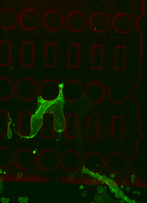
The clip was featured in a fascinating documentary on BBC Four that explores the scientist and broadcaster, Dr George McGavin’s diagnosis of the skin cancer melanoma. It shows melanoma cells moving easily through a maze of microscopic holes.
These mutant cells can pass through gaps smaller than their own nucleus, an amazing feat that gives them their deadly advantage. And they can use this technique to evade the immune system and hide from attack.
Catching cancer cells in the act of these rapid shape changes is key to understanding the mechanisms that control it, and if doctors could control or prevent it, they could stop the cells from spreading and make cancer more treatable.
Finding the genes behind cell shape
Professor Bakal and his lab at the ICR have been working for years to identify the genes and proteins that are involved in controlling cell shape.
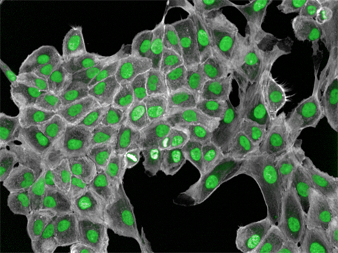
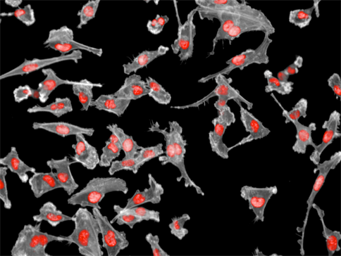
Images: Non-metastatic breast cells (top) vs metastatic breast cells (bottom)
Their work wouldn’t be possible without access to cutting-edge imaging technology as well as the computational methods and expertise needed to make sense of the huge amount of data produced.
Dr Bakal explained that every cell has about 20,000 genes that can produce millions of proteins. In cancer, hundreds or even thousands of these proteins will play a role in regulating cell shape, so you need to know where to look find the right ones that could be targeted to treat the disease.
And this is one of the key strengths of Professor Bakal’s lab – world class researchers with a deep knowledge of cancer biology who are also extremely skilled at working with cell imaging data to find that needle in a haystack.
Using large scale genetic screens and algorithms to recognise features of cells, they are gaining a unique understanding into what is happening in cancer cells when they metastasise.
Seeing proteins in super-resolution
Professor Bakal and his team have identified hundreds of proteins that play a role in the shape of cells in cancer:
“We’ve found interesting proteins, we’ve found interesting genes, now we want to find out what they’re doing.”
Physicist Eric Betzig at the Janelia Farm Research Campus in the USA won the 2014 Nobel Prize in Chemistry for his role in developing super-resolution microscopy. Professor Bakal has been working with super-resolution technology only available at Janelia Farm to look at cancer cells closer than ever before.
They are studying focal adhesions in cancer cells – groups of proteins that form finger-like attachments on the surface of cells that help them sense their environment.
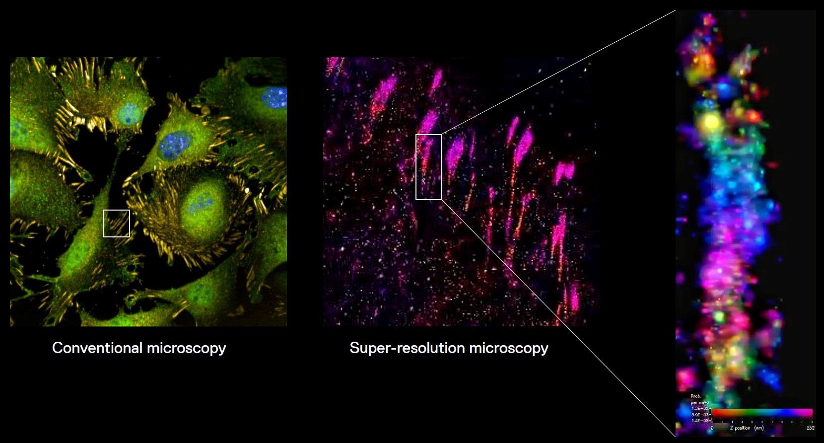
Image: Super-resolution microscopy images of focal adhesions, which can produce highly detailed images of regions of cells down to individual proteins, which is helping to understand more about processes like metastasis.
Using super-resolution microscopy, they have focused in on single focal adhesions on cancer cells to see how their proteins are arranged.
Looking at cancer cells in such unprecedented detail is key to understanding how their focal adhesions differ to those found on healthy cells, which could help explain how cancer cells move and spread.
“Were using super resolution microscopy to really see these structures in cancer cells for the first time,” said Professor Bakal. “No-one has ever seen this before because they haven’t been able to look at cells so closely.”
They hope their research will identify proteins that could be targeted by drugs to prevent cancer cells from changing shape and improve treatment.
Professor Bakal said: “If we can target cell shape changes, if we can freeze cancer cells in one particular shape, we can stop them from metastasising, and we can turn an incurable disease into a curable one.”
The next step for Professor Bakal’s lab is to combine the detail of super-resolution microscopy with the speed of other microscopy techniques. He’s doing just that with Imperial College London – the two institutions are working together to build a new high-speed super-resolution light sheet microscope.
This new microscope will be able to produce hundreds of ultra-detailed images of cells in just 30 minutes, compared with hours per image at the moment, which will really speed up what they are able to learn about cancer cells and metastasis.
Sharing his exciting findings, Professor Bakal emphasised that it takes more than the best technology to make the discoveries that defeat cancer, it also take world class researchers to make connections and ask the right questions, and that continued support is vital to keep pushing cancer research forward.
“Cutting-edge microscopy is essential to find out how to stop cancer cells from changing shape, but the best microscopes in the world need to be paired with new computational methods, artificial intelligence and big data methods to find new drugs.”