Photoacoustic Absorption Spectroscopy and Gold Nanorods for Nolecular Imaging
D Birtill, M Jaeger, A Shah, A Gertsch JC Bamber; in collaboration with E O’Flynn, N deSouza, Radiology Department and CRUK-EPSRC Cancer Imaging Centre; S Robinson, CRUK-EPSRC Cancer Imaging Centre;
Source of funding: Institute of Cancer Research, CRUK, EPSRC, EU FP6 Marie-Curie
Photoacoustic (PA) imaging is showing promise for visualising molecularly specific information associated with intrinsic chromophores such as oxyhaemoglobin and deoxyhaemoglobin, or external agents such as nanoparticles, which may be functionalised to bind to molecular targets of interest.
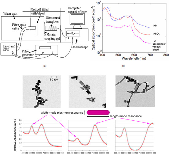
We have built a PA spectroscopy system (Figure 11a) to study the differences between the PA spectra from these media, with a view to optimising their identification in clinical multi-wavelength PA images. A variable-wavelength laser delivers short (ns) pulses of light, via a fibre optic cable, into a sample held between 75 µm transparent membranes. The optical wavelength is controlled by a computer, which scans the wavelength range 400-700 nm using a different pulse for each wavelength. Each pulse causes the sample to momentarily expand and emit a pressure wave, the energy of which is measured by the computer using a digital oscilloscope that samples the signal from a strongly focused ultrasound transducer. The resulting spectra are corrected for the wavelength-dependent laser energy. Further corrections are planned, so that the measurement is truly of optical absorption coefficient at each wavelength. Even without these additional corrections, the measured PA spectrum of oxygenated blood strongly resembles the published optical absorption spectrum (Figure 11b).
These results suggest that, in addition to its intended use for determining the optimum wavelengths for clinical PA imaging of blood oxygenation level and contrast agent concentration, this system may have applications as a laboratory spectrophotometer. Unlike traditional transmission spectrophotometers, which measure the extinction coefficient, the PA spectrometer will measure the absorption coefficient. This will make it suitable for use with (a) optically dark samples such as normal blood, which cannot be analysed in a standard spectrophotometer without dilution, and (b) turbid media, which normally require an optical scatter correction to convert the extinction coefficient to an absorption coefficient. Gold nanorods have also been synthesised in-house to various lengths (Figure 11c), with a view to investigating multiplexed PA molecularly-targeted imaging.
 Fig.11. (a) Photoacoustic absorption spectrometer. (b) Preliminary relative photoacoustic absorption spectrum of venous blood compared to standard data for haemoglobin and oxyhaemoglobin. (c) Transmission electron microscope images and corresponding optical extinction spectra for in-house synthesized gold nanorods.