Pinpointing the first founder mutations that lead to leukaemia

The ICR's Professor Mel Greaves and Professor Richard Houlston have identified mutations in the womb that lead to leukaemia.
In a study observing how leukaemias develop in pairs of identical twins, Professors Mel Greaves and Richard Houlston were able to identify the exact mutations that kick-start the cancer. By pinpointing the founder mutations driving the disease, they hope more effective targeted therapies that are less prone to drug resistance might be found.
Their team found identical twins in which both individuals had developed the blood cancer acute lymphoblastic leukaemia (ALL), the most common childhood cancer. By sequencing the whole genomes of leukaemic cells, they were able to draw up a list of the mutations that were driving the disease. Comparing the list of mutations for each twin, they could work out which were shared — suggesting that they were early founder mutations — and which had been acquired later.
The work may help us to tackle one of cancer's biggest challenges: drug resistance. Many cancer therapies target specific mutated genes that drive the disease. But as they progress, cancers evolve and diversify, with different cells picking up an ever more diverse set of different mutations. If a therapy is targeted to one of those later mutations, it will not destroy all of the cells, and so the disease will be able to return. The goal is to target the founder mutation, shared by all of the cancer cells. To use the analogy of the evolutionary tree, we must sever the trunk rather than prune the branches.
The twins study provided a unique opportunity to look for these founder mutations. In identical twins where one child develops leukaemia, it is relatively common (around 10% of twins) for the sibling to also develop the disease. This is because pre-leukaemic cells containing the founder mutation are able to pass between the twins in the womb, via the circulatory system that they share with the placenta. These clones then develop and evolve separately in the two individuals, picking up different sets of mutations, but retaining the original shared founder mutation.
References
Ma, Y., et al. (2013) Developmental timing of mutations revealed by whole-genome sequencing of twins with acute lymphoblastic leukemia. Proc Natl Acad Sci, 110:7429-7433 doi:10.1073/pnas.1221099110
Using virtual experiments to uncover missing cancer targets

Dr Bissan Al-lazakini and her team at the ICR have identified 46 previously overlooked but potentially 'druggable' cancer targets, using a powerful new online knowledgebase called CanSAR.
CanSAR was developed by The Institute of Cancer Research, London, with funding from Cancer Research UK, allowing researchers to carry out 'virtual experiments' to quickly prioritise which are the best targets for drug discovery.
While world-wide initiatives in cancer genomics are generating unprecedented volumes of cancer data, the translation of these genome data to drugs has been a slow and increasingly daunting process. This is mainly due to the amounts of information from different disciplines that need to be collated and examined to form this bridge between genome data and drug prototypes.
In response to this challenge, Dr Bissan Al-Lazikani and her team have created CanSAR – a cancer knowledgebase that integrates vast amounts complex data from patients, clinical trials and genetic, biochemical and pharmacological research. CanSAR scans these data and uses artificial intelligence, like that used in weather forecasts, to predict which potential drugs are likely to work in which circumstances.
Demonstrating the remarkable potential of CanSAR, the team analysed the Sanger Institute's existing list of 479 cancer genes. They found 46 potential protein targets that had until now been ignored despite their known biological relevance to cancer. These results show that, using this approach, we can find targets for cancer drugs in a smarter and faster way than ever before, paving the way for research into promising new drug targets that until recently may have been overlooked because of a lack of information. The analysis was carried out using the prototype of CanSAR, and a new version of the knowledgebase was launched in November this year. The new version now contains more than one billion experimentally derived measurements, nearly one million biologically active chemical compounds and data from over a thousand cancer cell lines. It also contains drug target information from the human genome and model organisms.
The clues needed to tackle cancer are hidden in data like this and by making it freely available to scientists across the globe, researchers can collaborate and make breakthroughs sooner. By selecting the very best targets that are most likely to lead to successful drugs, the success rate in the clinic increases. Not only will this speed up drug discovery and operate at a lower cost compared with traditional routes, but it will enhance innovation – shifting the focus away from tried and tested drug targets while managing the inevitable risk associated with moving into new and exciting areas. Both patients and the pharmaceutical industry will benefit tremendously from these advances.
References
Patel, MN., Halling-Brown, MD., Tym, JE., Workman, P. & Al-Lazikani, B. (2013) Objective assessment of cancer genes for drug discovery. Nat Rev Drug Discov, Vol.12(1), pp.35-50 doi:10.1038/nrd3913
Discovering an intriguing gene link to breast and ovarian cancer
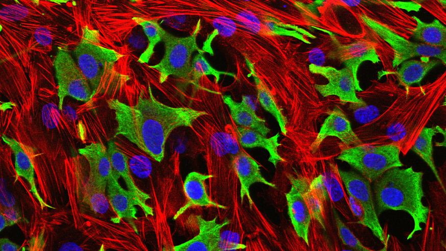
A team of researchers led from the ICR found that 'mosaic' mutations in a gene called PPM1D are linked to an increased risk of breast and ovarian cancer – through what may be a new mechanism of cancer development.
The PPM1D mutations were only found in blood cells from patients, and not in tumours themselves or in the normal breast or ovarian cells. It is possible that similar mosaic mutations – affecting some cells in the body but not others – will emerge in other genes, and in patients with other types of cancer.
Women with PPM1D mutations have a 20 per cent chance of developing breast or ovarian cancer – double the breast cancer risk and over 10 times the ovarian cancer risk of women in the general population.
The discovery could have implications for genetic testing and targeted prevention in particular for ovarian cancer, which is often diagnosed at an advanced stage.
And intriguingly, PPM1D seems to be working in a completely different way to other genes known to increase the risk of breast and ovarian cancer, such as BRCA1 and BRCA2.
We have two copies of every gene. In most cancer-causing genes a mutation in one copy is inherited and present in every cell, with the second copy mutated in the tumour itself.
However, in this case the team found PPM1D mutations were not inherited and rather than being present in every cell, were only found in blood cells.
The mutations make PPM1D overactive, which in turn reduces the action of a gene called TP53, which is the most frequently mutated gene in cancer cells.
Study leader Professor Nazneen Rahman, Head of Genetics at The Institute of Cancer Research (ICR) and of the Cancer Genetics Clinical Unit at The Royal Marsden NHS Foundation Trust, said: "This is one of our most interesting and exciting discoveries. At every stage the results were different from the accepted theories. We don't yet know exactly howPPM1D mutations are linked to breast and ovarian cancer, but this finding is stimulating radical new thoughts about the way genes and cancer can be related.
"The results could also be useful in the clinic, particularly for ovarian cancer which is often diagnosed at an advanced stage. If a woman knew she carried a PPM1D mutation and had a one in five chance of developing ovarian cancer, she might consider keyhole surgery to remove her ovaries after completing her family."
Revolutionary new sequencing technologies that allow much deeper analysis of genes were crucial to the discovery. The PPM1D mutations were only present in some cells – and this sort of mosaic pattern is difficult to detect with older sequencing methods.
The team analysed 507 genes involved in DNA repair in 1,150 women with breast or ovarian cancer, identifying PPM1D gene mutations in five women. They then sequenced the PPM1D gene in 7,781 women with breast or ovarian cancer and 5,861 people from the general population. There were 25 faults in the PPM1D gene in women with cancer and only one in the general population, a highly statistically significant difference.
References
Ruark, E., Snape, K., Humburg, P., Loveday, C., Bajrami, I., Brough, R., Rodrigues, DN., Renwick, A., Seal, S., Ramsay, E., et al. (2013) Mosaic PPM1D mutations are associated with predisposition to breast and ovarian cancer. Nature, Vol.493(7432), pp.406-U152 doi: 10.1038/nature11725
The study was funded by the ICR, the Wellcome Trust, Cancer Research UK and Breakthrough Breast Cancer.
Finding the faulty gene with a domino effect on cancer risk
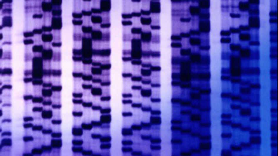
ICR scientists identified a brand new genetic mechanism that raises the risk of cancer via a domino effect on DNA – with one gene fault triggering a second, cancer-causing mutation.
Professors Richard Houlston, Gareth Morgan and their team showed that patients with a mutation in the CCND1 gene were 80 per cent more likely to develop a particular sub-type of myeloma, a blood cancer, than those without the mutation.
The sub-type is known to be triggered by a specific chromosomal translocation – a type of DNA breakage and rearrangement – designated by the code t(11;14). This sub-type, which affects about a fifth of myeloma patients, involves the translocation forming a mutant piece of DNA that drives cancer by activating several genes. The study was the first time a genetic mutation has been linked to a specific chromosomal mutation.
The study assessed the DNA of 1,655 patients with myeloma and compared them with people in the general population, in order to explore whether they had inherited genetic faults that had increased their risk of developing the disease.
Researchers hailed the finding as a “very important discovery” that opens up new avenues for cancer treatment and prevention.
Professor Richard Houlston, Professor of Molecular and Population Genetics at the ICR, said: “We’ve uncovered a new way in which genetics can raise the risk of developing cancer, in which one gene fault appears to trigger another – in a domino-like effect.
“It’s the first time anyone has identified a specific gene fault that raises the risk of developing a second specific type of DNA damage. Not only is the discovery valuable for our understanding of multiple myeloma, but it also demonstrates the value of studying specific genetic sub-types of cancer, rather than grouping them all together as one disease.”
Professor Gareth Morgan, Head of Molecular Haematology at the ICR and Head of the Myeloma Unit at The Royal Marsden, said: “This is a very important discovery, which opens out our understanding of cancer genetics and gives us new avenues for treatment and prevention.
“Now that we have found a genetic risk factor for a particular sub-type of multiple myeloma, I’m hopeful we can design more targeted treatments for the people who develop it. In cancer research we tend to group different sub-types together, but this research proves we need to move away from this approach and investigate sub-types at a molecular level.”
References
Weinhold, N., Johnson, DC., Chubb, D., Broderick, P., Goldschmidt, H., Hemminki, K., Foersti, A., B, C., Hosking, F., Ma, Y., et al. Vogan, K., (2013) The CCND1 G870A polymorphism is a risk factor for t(11;14)(q13;q32) multiple myeloma. Nature Genetics, pp.522-U87 doi:10.1038/ng.2583
The study was funded by a number of organisations, including Myeloma UK, Leukaemia & Lymphoma Research, Cancer Research UK and via the NIHR Biomedical Research Centre at The Royal Marsden NHS Foundation Trust and The Institute of Cancer Research.
ICR's pioneering prostate cancer drug found to be effective before chemotherapy

Abiraterone, a prostate cancer drug discovered at the ICR, showed impressive benefits in a major clinical trial of men with early-stage prostate cancer.
The drug has already been approved by NICE for men with advanced prostate cancer, but could now benefit men with earlier disease who have not yet received chemotherapy.
The phase III trial, carried out with the ICR and The Royal Marsden NHS Foundation Trust, showed that when the drug was given to men with no or mild symptoms before chemotherapy it could double the time before tumour progression could be detected by a scan.
Professor Johann de Bono and colleagues gave abiraterone to 1,088 men with prostate cancer that no longer responded to standard hormone-suppressing drugs but who had not received chemotherapy.
Men taking abiraterone went an average of 16.5 months before tumour growth was detected by a CT or MRI scan – twice as long as the 8.3 months among men taking a placebo. Men in the abiraterone group also reported less pain than those on placebo.
The benefits seen in some men were so impressive that the trial was stopped early so that all men in the study could receive it.
Men with advanced prostate cancer are already seeing the benefits of abiraterone, with patients living longer and suffering fewer side effects. The trial’s findings, which were published in the New England Journal of Medicine, pave the way for more men with prostate cancer to be treated with abiraterone earlier in the course of their disease, without having to undergo toxic chemotherapy first.
Abiraterone was discovered at the ICR in what is now the Cancer Research UK Cancer Therapeutics Unit and further developed at the ICR and The Royal Marsden.
References
CJ Ryan, MR Smith, JS de Bono, A Molina, CJ Logothetis, P de Souza, et al,(2013) Abiraterone in metastatic prostate cancer without previous chemotherapy. New England Journal of Medicine 368 (2), 138-148 DOI: 10.1056/NEJMoa1209096
Gene mutation drives childhood glioblastoma
.jpg?sfvrsn=5b267169_0)
An ICR research team led by Dr Chris Jones uncovered the genetic causes of a rare but lethal form of childhood brain cancer, improving our understanding of the development of the disease.
The team discovered a mechanism which drives the development of childhood glioblastoma, opening up the prospect of an effective treatment for the disease for the first time.
About 60 children develop childhood glioblastoma in the UK each year, and no drug treatments are currently available. Glioblastoma is more than one disease, with different types developing at different ages and in different areas of the brain. One type affects younger children aged from about six years and another affects children and young adults aged 13 and over.
The different forms are characterised by different mutations in a histone protein, H3F3A - a gene scaffold protein which controls the activity of other genes. The ICR researchers looked at datasets from patients with the different forms of the disease and showed that the different types of glioblastoma affecting older and younger children had distinct patterns of gene activity.
To investigate the significance of these different histone mutations the team looked at cells taken from a glioblastoma patient with the form of the disease affecting older children, to look at the expression of other genes which may be driven by the mutated form of the histone protein. They discovered that in the cells with this mutation, a potent oncogene (cancer-causing gene) called MYCN was highly active.
Once they found out that this mechanism was involved in the development of this form of glioblastoma, the team ran a large-scale screening experiment to see what might be a good target in blocking the cancer-causing effect of the MYCN gene. They identified a drug target called aurora kinase A, for which drugs are already being developed.
These drugs can now be tested in the particular group of children with glioblastoma who have the histone mutation, and the researchers plan to test these in clinical trials of children within two to three years. Hopefully this opens up the prospect of an effective treatment for the disease for the first time.
References
Bjerke, L., Mackay, A., Nandhabalan, M., Burford, A., Jury, A., Popov, S., Bax, DA., Carvalho, D., Taylor, KR., Vinci, M., et al. (2013) Histone H3.3 Mutations Drive Pediatric Glioblastoma through Upregulation of MYCN. Cancer Discovery 3(5), pp.512-519 doi: 10.1158/2159-8290.CD-12-0426
Unlocking the hidden potential of cutting-edge cancer drugs

In a collaboration with the University of Sussex, Professor Paul Workman and his team at the ICR uncovered a hidden mechanism revealing how a class of cutting-edge cancer drugs called protein kinase inhibitors attack tumours, which they hope could keep patients alive for longer.
Protein kinase inhibitors are some of the most heralded of the new kind of targeted therapies, with 25 already in use and around 400 under development. They treat 5,000-10,000 patients in the UK each year, with that number set to grow as more are approved for use. Kinase inhibitors work across types of breast, skin, lung, kidney and other cancers, but while effective often only extend life by around three to six months. After uncovering the previously hidden mechanism of action, scientists believe they can unlock the true potential of the drugs by changing the way they are used.
It had been thought that the drugs work solely by blocking the cell signalling function of enzymes called protein kinases – which play an active role in many cancers – by preventing them from binding to ATP, the basic unit of energy in cells. But this new major discovery has found that at high doses, the drugs also block kinases from linking up with a complex of molecules in cells called the Hsp90-Cdc37 chaperone system, which is essential for maintaining the stability of proteins. The team showed that this 'chaperone deprivation' actually caused the destruction of the cancer-causing kinases and halted the growth and division of cancer cells.
The researchers now plan to conduct clinical trials using kinase inhibitors at higher doses but with rest periods to take advantage of the new mechanism – and believe the new method has the potential to keep cancers at bay for much longer.
Professor Paul Workman, Chief Executive of the ICR, said: "We hope to carry out clinical trials to test the benefits of delivering kinase inhibitors in a way that should maximise their impact in destroying their targets. There is more work to do to prove the benefit to patients, but these drugs are already approved so there are fewer regulatory burdens than usual to overcome to test our new idea. Our results also support ongoing and future clinical trials in which we combine protein kinase drugs with HSP90 inhibitors – such as the drug we discovered AUY922 – to maximize the chaperone deprivation effect."
References
Polier, S., Samant, RS., Clarke, PA., Workman, P., Prodromou, C. & Pearl, LH. (2013) ATP-competitive inhibitors block protein kinase recruitment to the Hsp90-Cdc37 system. Nat Chem Biol, Vol.9(5), pp.307 doi:10.1038/nchembio.1212
The study was funded by Cancer Research UK and the Wellcome Trust.
Beating breast cancer’s resistance to hormone treatments

ICR scientists uncovered a mechanism by which breast cancers can develop resistance to an important class of drugs called aromatase inhibitors used in the hormonal treatment of the disease.
The findings could be used to develop drugs that prevent breast cancers from building resistance to aromatase inhibitors, allowing more women to benefit from hormone therapy.
Aromatase inhibitors are an important part of hormone treatment for oestrogen receptor (ER)-positive breast cancer, which accounts for 70% of all newly diagnosed cases of breast cancer.
These tumours need oestrogen to survive and grow, so drugs called aromatase inhibitors, which can stop women’s bodies from producing oestrogen, are used in their treatment.
A study led by Professor Clare Isacke at the ICR discovered that the GDNF-RET signalling pathway in ER-positive breast cancer is linked to resistance to aromatase inhibitors in some cancers.
Her team found that breast cancer cells which have developed resistance to the drugs have higher levels of signalling by the GDNF-RET pathway than cells without resistance to aromatase inhibitors. ER receptors in the resistant cells were activated by GDNF-RET pathway signals, allowing tumours to grow even when treatment was blocking oestrogen production. But using a drug to block GDNF-RET signalling allowed these cells to become sensitive to treatment with aromatase inhibitors once again.
Breast cancer patients whose tumours had an activated GDNF-RET pathway were more likely to develop resistance to treatment with aromatase inhibitors than those whose pathway was not activated. They also had an increased risk of relapses and worse long- term chances of survival than patients whose tumours did not activate the GDNF-RET pathway.
Professor Isacke and her team hope the findings will help doctors to predict response to aromatase inhibitors and pick out who will benefit most from the treatment. Having discovered the importance of the GDNF-RET pathway in forming resistance to hormone therapy, the researchers believe that switching off this pathway in the clinic will increase the effectiveness of aromatase inhibitor treatment for women with breast cancer.
References
Morandi, A., Martin, LA., Gao, Q., Pancholi, S., Mackay, A., Robertson, D., Zvelebil, M., Dowsett, M., Plaza-Menacho, I. & Isacke, CM. (2013) GDNF-RET signaling in ER-positive breast cancers is a key determinant of response and resistance to aromatase inhibitors. Cancer Research 73:3783-3795 doi: 10.1158/0008-5472.CAN-12-4265
World’s largest study quantifying breast cancer risk after treatment for Hodgkin lymphoma
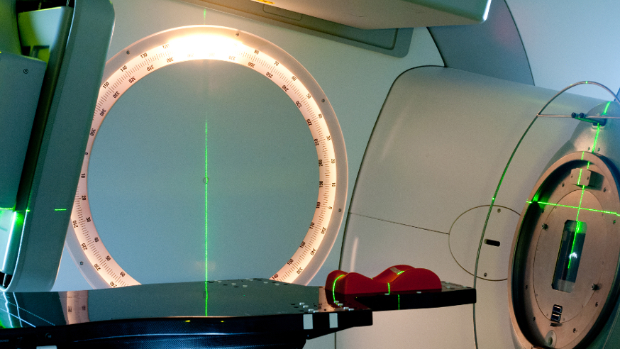
In the world’s largest cohort study, The ICR’s Professor Tony Swerdlow and colleagues investigated factors affecting the risk of developing breast cancer after radiotherapy treatment for Hodgkin lymphoma.
Hodgkin lymphoma is a disease of the lymphatic system, where cells in the lymph nodes become cancerous, and is commonly treated with radiotherapy or chemotherapy. It has been known since the 1990s that treating Hodgkin lymphoma with radiotherapy is associated with an increased risk of breast cancer later in life.
Professor Tony Swerdlow and his team analysed data from 5,000 women who had been treated with a treatment called supradiaphragmatic radiotherapy (radiotherapy performed above the diaphragm) between 1956 and 2003 to determine breast cancer risk in relation to their treatment.
The team looked at factors such as patient age at treatment, the type of treatments they received, and how long ago they received treatment. Combining this information with breast cancer occurrence, the team could calculate relative breast cancer risk for a number of different scenarios by comparing breast cancer incidence in the different groups against the general population rate of breast cancer. Because of the large number of women included in the study, the risk calculations had greater precision than those previously.
Most excitingly, the team were able to use these results to describe cumulative risks for different types of patient, bringing together the different scenarios of an individual’s treatment. This can be used to calculate an individualised estimate of risk for patients, providing information for personalising follow-up and preventive measures according to treatment type, age, and time.
The findings suggest some aspects of treatment and follow-up that need to be considered to potentially decrease the chances of a patient with Hodgkin lymphoma developing breast cancer later in life or to detect the disease early. For example, the analysis suggests that more intensive breast cancer screening programmes could be used for certain women at particularly high risk of breast cancer.
The study found that patients who were treated with radiotherapy around the age of puberty were at greater risk of breast cancer than those who were treated later in life and points to the need to consider whether special measures should be undertaken to reduce the risk for girls undergoing radiotherapy at these ages.
This large cohort study has helped identify risk factors for breast cancer for patients with Hodgkin lymphoma, and gives a useful underpinning for personalising treatments and patient follow-up and therefore, it is hoped, leading to improved outcomes for patients.
References
Cooke, R., Jones, ME., Cunningham, D., Falk, SJ., Gilson, D., Hancock, BW., Harris, SJ., Horwich, A., Hoskin, PJ., Illidge, T., et al. (2013) Breast cancer risk following Hodgkin lymphoma radiotherapy in relation to menstrual and reproductive factors. Br J Cancer, Vol.108(11), pp.2399-2406 doi:10.1038/bjc.2013.219
Stopping cancer in its tracks
.jpg?sfvrsn=aa459a68_2&Width=874&Height=492&ScaleUp=false&Quality=High&Method=CropCropArguments&Signature=7CCCCF64AA95BB10266B7FE9839FAA9C)
Dr Chris Bakal and his team at the ICR have identified mechanisms cancer cells use to 'shape-shift' to invade different types of tissue, which could help develop treatments for aggressive skin cancer.
Researchers know cancer cells have to take on different shapes so they can metastasise, or move around the body. For example, in the blood and in soft tissues, cancer cells adopt a rounded shape. But when travelling across bone and other hard tissues, they become elongated. It’s like us getting across ice and water – we need different pieces of equipment to do this. Without the ability to assume these shapes, cancer cells cannot spread.
Until recently, we knew hardly anything about the mechanisms behind this shape switch. But Dr Chris Bakal and his team have pinpointed a set of genes that allows melanoma cells to change rapidly between two shapes in order to escape from the skin and spread around the body.
Researchers saw that by switching off a gene called PTEN, they could increase the number of elongated cells, and this appears to help the disease move through tissues. PTEN is an important tumour suppressor – a gene that appears to stop healthy cells from becoming cancerous – and it is switched off in around one in eight patients with melanoma and in almost half of those who carry a mutation in another cancer gene called BRAF.
Scientists think one way melanoma cells can metastasise is by losing their PTEN function, increasing their shape-shifting ability and in turn enabling them to move to many different tissues within the body.
These early findings could pave the way for scientists to develop desperately needed drugs for malignant melanoma which kills more than 2,200 people every year. Melanoma spreads very easily to other areas like the liver, lungs or brain, and once it has spread it becomes very difficult to treat. Surgery or radiotherapy may eradicate tumours at their site of origin, but current treatment options are rarely able to cure cancer once it has spread. Crucially, by understanding how cells shape shift, we might find a way to control metastatic cancer through drugs or other therapies.
References
Zheng Yin, Amine Sadok, Heba Sailem, Afshan McCarthy, Xiaofeng Xia, Fuhai Li, Mar Arias Garcia, Louise Evans, Alexis R. Barr, Norbert Perrimon, Christopher J. Marshall, Stephen T. C. Wong & Chris Bakal. A screen for morphological complexity identifies regulators of switch-like transitions between discrete cell shapes. Nature Cell Biology (2013) doi:10.1038/ncb2764